
The fluorescence spectroscopy of chlorophylls in plants and algae is an effective non-invasive technique to assess their physiological state and photosynthetic activity. Changes of the fluorescence yield and kinetics indicate the disruption of photosynthesis and metabolic processes. Thus, autofluorescence measurements can be used for toxicity assessment[1]. Heavy metal-containing quantum dots (QDs) represent nanoparticles that are also useful as luminescent markers for biological imaging owing to the enhanced photostability, bright tuneable photoluminescence (PL) and wide possibilities for surface modification.
In this work the fluorescence spectroscopy and microscopy methods were applied to evaluate the effects of different concentrations of copper ions (Cu2+) and water soluble CdTe-MSA quantum dots (a peak of photoluminescence at 550 nm) on the green freshwater Chlorella sp. and Scenedesmus sp. microalgae. When exposed to blue light, chlorophylls of healthy microalgae cells fluoresce brightly in the red spectral region as detected by measuring autofluorescence spectra by means of a spectrophotometer Perkin Elmer LS 55. Samples were kept under natural daylight conditions and remained in the dark overnight for adaptation before measuring the slow part of autofluorescence kinetics (at 683 nm, the peak of the main fluorescence band) using Ocean Optics USB 2000+ spectrometer (excitation wavelength – 405 nm).
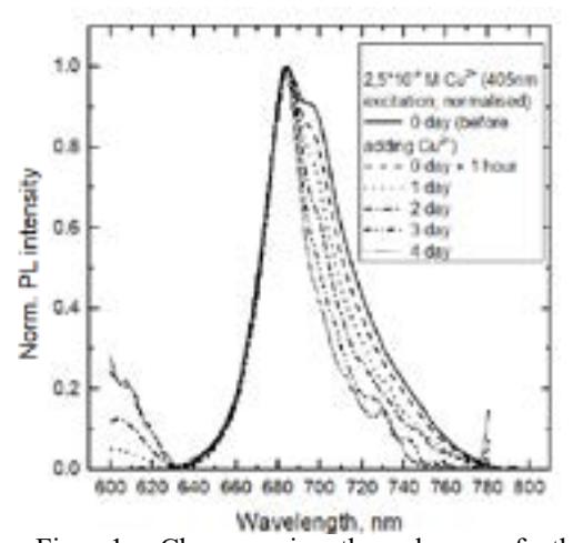
Changes in both intensity and form of the microalgae fluorescence spectra were observed when being affected by various concentrations of copper ions, in comparison to the control samples. The autofluorescence decreased depending on the ion concentration and the exposure time, indicating damage to the photosynthetic apparatus of microalgae. Regarding the changes in the spectral shape, the intensity of the longer-wavelength slope has decreased (Fig. 1).
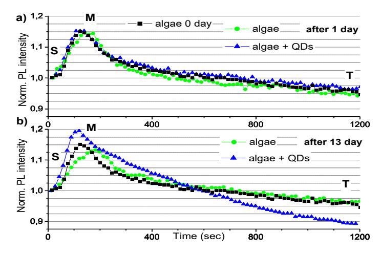
The pattern of the photoinduced fluorescence kinetics in microalgae consisted of the steady-state level "S", the maximum level “M” and the terminal level “T” (Fig. 2). The influence of QDs on the autofluorescence of algae cells became more pronounced on later days, and the biggest changes in the kinetics curve were observed after 13 days incubation with QDs, which included relative enhancement and further shift of the maximum "M" position as well as steeper FL intensity reduction from the level “M” to “T”.
The microscopic measurements were done by means of a fluorescence microscope (Nikon eclipse 80i). There were no obvious changes seen in algae cells using a phase contrast mode after 24 hours of incubation with copper ions or QDs. However, fluorescence images, taken using UV or violet spectral excitation, showed some of round-shaped Chlorella sp. algae starting to photoluminesce in yellow in the samples with QDs, and the red fluorescence intensity in green algae cells was disappearing in a non-uniform manner in the presence of copper ions in comparison with those in the samples of microalgae kept without heavy metals. The contact with QDs resulted in decrease of the autofluorescence starting from the second day of incubation, which seems to be related with the reduced quantum yield of chlorophylls and does not involve the direct inactivation of algae cells during the first week of incubation.