
Electronic properties and function of graphene depend on the surface structure, origin of defects and adsorption of molecules at surface. Riboflavin can be employed in electrochemistry for different purposes as well as it can be detected electrochemically due to its aromatic nature. Riboflavin has been used as an electrode modifier in sensor construction for other important compounds such as iodate, hydrogen peroxide, persulphate [1].
Graphene science and development of graphene-based technologies requires to control adsorption processes and obtain molecular level knowledge of surface. To understand surface and interfacial chemistry, sensitive spectroscopic technique is needed. Surface-enhanced Raman spectroscopy (SERS) is one of the most sensitive surface analysis technique and fulfils such requirements. Unfortunately, SERS is limited by certain substrates (mostly Ag, Au, and Cu) and requirement to use roughened/nanostructured surface restrict the applicability of this method. Consequently, Tian et al. approached a novel SERS technique named "shell-isolated nanoparticle-enhanced Raman spectroscopy" (SHINERS). The method is based on enhancement of Raman signal by strong electromagnetic field provided by gold core nanoparticles surrounded by a few nanometer thick inert silica shell (Au@SiO2) [2]. Recently, we employed SHINERS for analysis of monolayer at smooth gold Au in situ at controlled potential [3]. In this study, SHINERS was used to probe the structure of adsorbed riboflavin at graphene layers at molecular level.
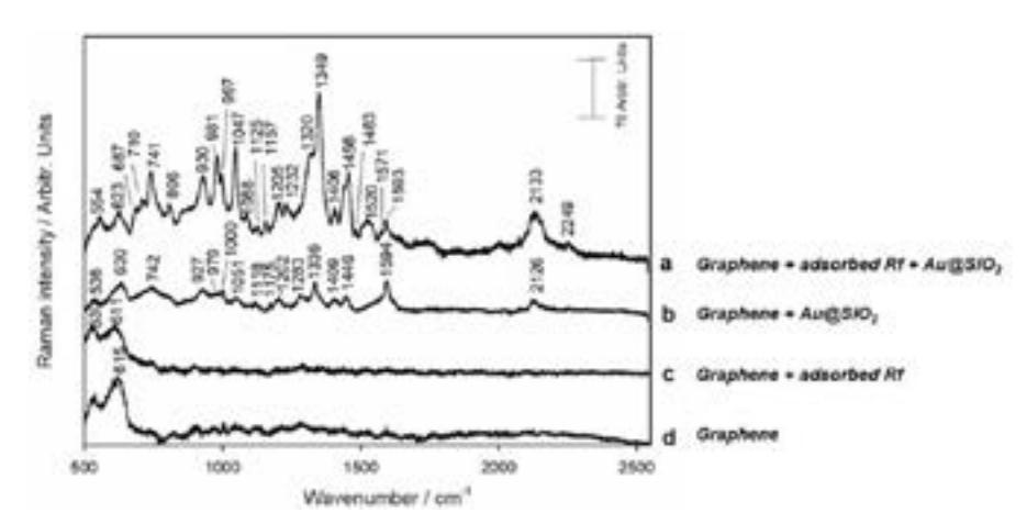
It has been determined that bands from copper oxide, carbon network, defect sites and adsorbed riboflavin (Rf) are visible in SHINERS spectra. It allowed us to obtain all riboflavin characterized vibrational modes. To conclude, by adsorbing riboflavin molecules at surfaces of graphene, we have shown that SHINERS spectroscopy provides possibility to probe any adsorbed molecules at graphene.