
Size distribution of noble metal nanoparticles strongly influences their optical properties [1]. In most of the cases linear dimensions and shape of the nanoparticles is determined by the applied synthesis method and after synthesis it no longer can be varied. However, irradiation with ultrashort laser pulses can be employed to tailor the size distribution of nanoparticles even after their fabrication [2]. In this work we have investigated how the morphology and size distribution of silver (Ag) nanoparticles embedded in diamond-like carbon (DLC) matrix – DLC:Ag nanocomposite thin films – deposited on silicon substrate [3] is altered after irradiation with the interference field of two Yb:KWG femtosecond laser beams (290 fs pulse duration, 2H 515 nm wavelength, 40 kHz repetition rate) [4]. Pristine and laser processed samples were investigated employing scanning electron microscopy (SEM). The size and distribution of silver nanoparticles in thin film was determined from SEM micrographs with 'ImageJ' software. Since the micrographs depicts micrometer range periodic patterns in films, further data filtering was applied to extract changes only in the laser affected areas. From the energy dispersive X-ray spectroscopy measurements it was determined that the investigated sample has 7.8 at.% silver content.
From the image analysis of the laser affected samples, average roundness of the nanoparticles was obtained to be 0.72 (perfect roundness is 1), thus in this particular case it was chosen to analyze area but not the diameter of nanoparticles. From the SEM micrographs of unaffected DLC:Ag films it was determined that an average area of nanoparticles was 530 nm2. Upon irradiation with fluence of 6 mJ/cm2 and 27k, 64k, 125k pulses, Ag nanoparticles with an average area of 1484, 854, 802 nm2 were obtained, respectively. From 600 to 1200 nanoparticles were uses in the analysis of the single SEM micrograph. Nanoparticles size distribution histogram in Fig. 1 depicts the particles area size distribution prior and after laser irradiation under different laser processing conditions
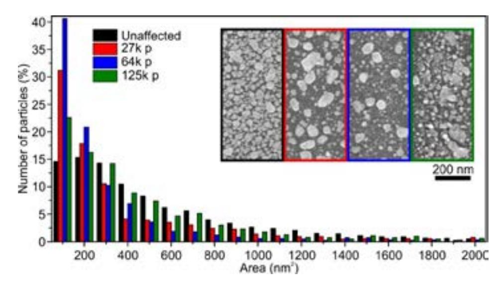
The image analysis revealed that increasing number of applied laser pulses affects on the average nanoparticle area which tends to decrease and at the same time increases the total number of nanoparticles. The later indicates fragmentation of Ag nanoparticles that results in increasing number of smallest particles after laser irradiation.