
Argonaute proteins (Agos) are widespread in all three domains of life (bacteria, archaea and eukaryotes), and are structurally highly conserved [1]. In eukaryotic organisms, eAgos constitute the functional core of the RNA-silencing machinery, which is critical for regulation of gene expression, silencing of mobile genome elements, and defence against viruses. According to the latest experimental data prokaryotic Agos (pAgos) constitute an additional defence system with high versatility against invading nucleic acids [1].
The structural organization of full-length pAgos, as well as eAgos is bilobal, composed of four domains. The N-terminal and the PIWI/Argonaute/Zwille (PAZ) domains together with the L1 and L2 linker regions constitute the N-terminal lobe, whereas the C-terminal lobe is composed of the MID and the P-element-induced wimpy testis (PIWI) domains, the latter harbouring the catalytic site of cleavage-active Agos (Figure 1).
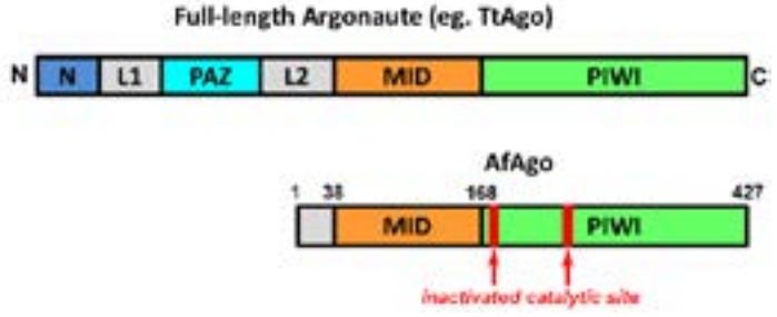
pAgos are further divided into two major groups termed long (full-length, as above) and short pAgos that lack the PAZ domain, the N-terminal domain and consequently the L1 linker region. The short Argonaute protein from an archaeon Archaeoglobus fulgidus (AfAgo) is composed of only the L2 linker region and the MID and PIWI domains and therefore corresponds to the "MID/PIWI" lobe of full-length Agos (Figure 1). Its PIWI domain is inactivated by mutations of active site residues. AfAgo is well crystallographically characterized with solved crystal structures of the apo protein and its complexes with RNA and DNA duplexes providing initial information on the molecular mechanism of RNA interference (RNAi) in eukaryotes [2-5].
Target recognition by Agos is realized via complementarity between the Ago-bound guide and the target strands (RNA or DNA). The 5'-end of the guide strand is anchored in the evolutionarily conserved pocked of the MID domain. Eukaryotic and prokaryotic Agos usually show a preference for a specific 5'-nucleotide of the guide strand (e.g. human Ago2 for a 5'-U, while bacterial TtAgo for a 5'-dC). Here we present biochemical and structural studies of AfAgo specificity for the 5'-end of the guide strand.