
Biologically modified surfaces, which employ a biomaterial, permit the development of biosensors. This analytical device consists of two main components: a biorecognition element and a transducer [1]. Human body has one of the most advanced recognition systems – an immune system. Immunosensor is a biosensor, in which an antibody or an antigen serves as the biorecognition element [2]. One of the biggest problems in creating immunosensors is the immobilization of the biomolecules [3]. Not all immobilization methods provide stability of biomolecules on the surface and site-directed positioning.
In order to evaluate the efficiency of the antibody-enzyme conjugate immobilization on the surface, scanning electrochemical microscopy (SECM) was employed. SECM is a non-invasive technique and can be used in an optimal medium for the biorecognition element. The aforementioned method provides the possibility to detect antibodies at localized surface areas and to determine the changes in electrochemical activity, by adjusting substrate and mediator concentrations in medium (fig. 1).
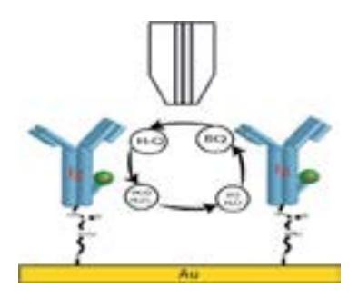
This study was conducted by using horseradish peroxidase labeled antibodies as biorecognition element, the red-ox mediator hydroquinone (H2Q) and hydrogen peroxide. Experiments were performed with three different mediators: ferrocene methanol, ferrocene carboxylic acid and hydroquinone, which proved to be the most effective red-ox mediator for the SECM surface imaging. The current measured with H2Q was about 3 times higher than with ferrocene carboxylic acid.
Therefore, SECM proved to be a suitable technique for validating the quality of the antibody immobilization and the enzyme's activity.