A success of nanotechnology in field of physical, chemical, and medical sciences, it has now started revolutionizing the drug delivery sciences and bio-detection. The specific advantages include superior pharmacodynamics, pharmacokinetics, reduced toxicity, and targeting capability.
It is well-known that hydroxyapatite (Ca10(PO4)6 (OH)2) is a form of bioceramics material and is widely used as a bone substitute due to its adequate mechanical properties and the similar composition to bone mineral. Due to the bioactivity, biocompatibility, stability, nontoxic properties, hydroxyapatite with porous surface structure and OH groups may serve as an ideal candidate drug carrier for the delivery of a variety of pharmaceutical molecules.
Metals such as gold, copper, europium or even ionic compounds of selenium are well-known for their potential anticancer properties. The synergy of such metals could bring promising effect by complete cancer cells eradication from healthy tissue.
The hexagonal structure in apatites belongs to $P6_3/m$ space group and allows the cations to localize in two different crystallographic positions and are able to accommodate a variety of cations as substituents and could find perfect application in bone cancer therapy.
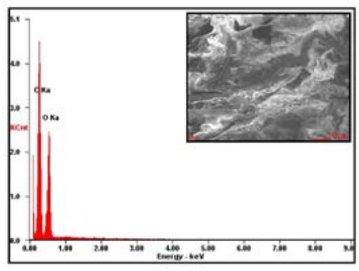
In the research there have been obtained porous scaffolds based on poly(L-lactide) (PLLA) and synthetic hydroxyapatite (HAp) co-doped with Sr2+, Eu3+ and Cu2+ ions using thermal induced phase separation technique (TIPS) supported by salt leaching process (SL). The obtained series of composite sponges consist of 50 wt.% of the hydroxyapatite in PLLA/HAp systems. The structural and morphological properties of the obtained samples were determined by using XRD (X-ray powder diffraction), TEM (transmission electron microscopy) and SEM (scanning electron microscopy) techniques (Fig. 1). In the present study, experimental in vitro anticancer tests concerning sponge scaffolds, which may substitute bone tissue, were discussed in detail.