
The volume and complexity of diagnostic imaging is increasing at a pace faster than the availability of human expertise to interpret it [1, 2]. Ophthalmic imaging provides a way to diagnose and objectively assess the progression of a number of pathologies. Automated diagnosis of a medical image, even for a single disease, faces two main challenges: technical variations in the imaging process, and patient-to-patient variability in pathological manifestations of disease. As far as we are aware, the state-of-the-art approaches tried to deal with combinations of these variations using an end-to-end black-box network, thus typically requiring quite a few of labeled samples. By contrast, our framework separates the two problems (technical variations in the imaging process and pathology variants) and deals with them independently. A segmentation network (uses modified U-Net architecture [3]) creates a detailed device-independent segmentation map. Subsequently, a deep classification network analyses this segmentation map and provides diagnoses and as the primary outcome one of four referral suggestions currently used in clinical practice.
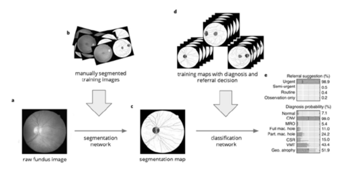
This study is focused on Diabetic Retinopathy and Glaucoma diseases. Our key insight is the ability to analyse simple fundus images that can be obtained through phones equipped with special lenses or simple fundus cameras and not be far from state-of-the-art approaches [4]. The method was tested on Kaggle Diabetic Retinopathy challenge, ORIGA, SCES, EyePACS-1 datasets, which were laid out in free access.
In conclusion, we present a novel framework that analyses clinical fundus images and makes referral suggestions to a standard that is comparable to clinical experts. Although we focused on one common type of medical imaging, future work can address a much wider range of medical imaging techniques, and incorporate clinical diagnoses. For future work, we consider the exploration of possible performance gains in other downstream tasks such as image level recognition or grading of diabetic retinopathy. Our method can be applied for other medical images as well. We can use AI to predict adverse outcomes before they happen, better manage highly complex situations, and ultimately allow clinicians to spend less time analysing data and more time harnessing their experience and human touch in delivering care.