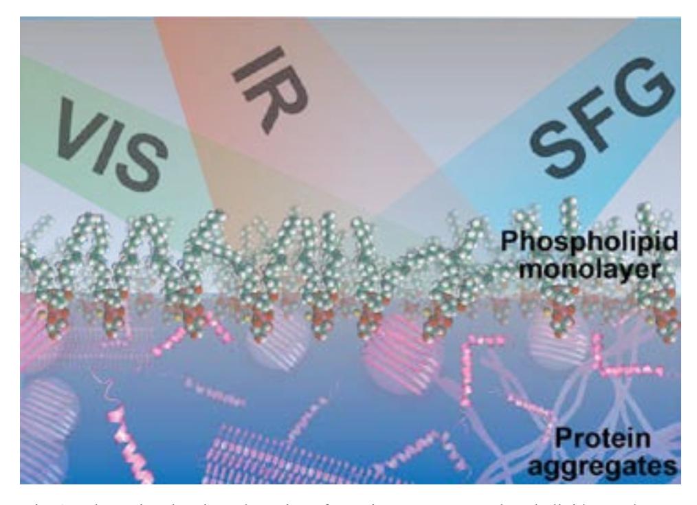
Formation of amyloid fibrils in cells and an intercellular net is associated with more than 40 different clinical conditions: Alzheimer's disease, Parkinson's disease, Huntington's disease, type II diabetes, and others. Each of these diseases caused by an aggregation of a different protein, which is a normal product of cellular metabolism. Protein elimination processes operating in parallel usually counterbalance production of the protein [1]. Nevertheless, when these production and elimination processes are disbalanced, protein starts to form different aggregates: small soluble oligomers and insoluble polymeric fibrils. All these aggregates are accumulating in tissues, causing inflammatory processes, tissue degradation and cell death.
The aim of our work is fully understand the protein aggregation process on a molecular level, to identify secondary structures of the aggregates and to investigate the interaction with phospholipid monolayer. It is believed that the interaction between protein and phospholipid layer greatly affects the aggregation process, therefore, it is important to understand how the molecular structure of the protein and its aggregates changes due to interaction with the lipid membrane. For this, a surface-sensitive spectroscopic method is needed. Vibrational sum-frequency generation spectroscopy is one of the most suited techniques to study protein aggregation and its interaction with phospholipid layer.
Vibrational sum-frequency generation (VSFG) spectroscopy is a surface sensitive and molecular specific technique. It is a widely used non-linear second order optical spectroscopic tool to study the conformation and orientation of proteins at various interfaces. Two pulsed laser beams, one of fixed visible frequency and the other of tunable infrared frequency overlap spatially and temporally at an interface to generate SFG signal (schematics shown in Fig.1.). The frequency of the generated SFG signal is the sum of the two incident field frequencies.
We applied the VSFG technique to study the structure of hen egg white lysozyme (HEWL) aggregates adsorbed at phospholipid monolayer. HEWL is an ideal model protein to study the mechanism of amyloid fibril formation. It has been studied extensively with other techniques and is closely related to human lysozyme, which also forms fibrils and causes hereditary systemic amyloidosis [2]. HEWL starts to form aggregates when the native structure of the protein is disrupted. This can be achieved by lowering the pH of the protein solution and elevating the temperature close to the protein's denaturation temperature. The protein aggregates adsorbed at the phospholipid monolayer due to a strong electrostatic interaction: negatively charged phospholipid monolayer attracted positively charged lysozyme aggregates. Whereas at the water/air interface only non-aggregated protein could be adsorbed. VSFG spectra were recorded in the Amide I region. The secondary structures of the aggregates were identified from the position and shape of this Amide I band.