
Scintillating detectors are popular as a means of detecting and measuring ionizing radiation. Due to their high stopping power, high light yield and impressive signal dynamic time inorganic scintillators have allowed for rapid progress in nuclear instrumentation in the last two decades. In recent times production of scintillation materials has become a limiting factor for future development in this field. While growing crystals from a melt allow for high crystal quality this method is limited by the maximum size of the crystal and variety of compositions.
Using glass and glass ceramic production methods allows for a more flexibility of crystal compositions and geometries. However, glass based scintillators have low light yields when excited with ionizing radiation. Using polycrystalline, glass-ceramic, materials would allow for advantages of crystalline materials to be combined with the flexibility of glass scintillators.
In out study we investigate three samples of scintillating grass-ceramics. Compostion of the investigated samples was $Gd_{1,485}Y_{1,485}Ce_{0,03}Al_2Ga_3O_{12}$ (GYAGG:Ce). Samples were sintered in identical temperature of 1650 °C. Two methods of pressing were used, ultrasonic and uniaxial. Sintering was performed with and without bed powders. After sintering hydrostatic density measurements revealed densities of 98.4%, 98.8% and 99,2% compared to the monocrystaline form. The samples were produced in National Research Center “Kurchatov Institute”, Moscow, Russia.
The study was performed by using confocal photoluminescence (PL) spectroscopy. A WITec Alpha300 S microscopic system coupled to a spectrometer with a thermally cooled CCD camera was used. A CW laser diode emitting at 405 nm was used for excitation. The spatial resolution of 250 nm in plane and 1 μm perpendicularly to the sample surface was achieved.
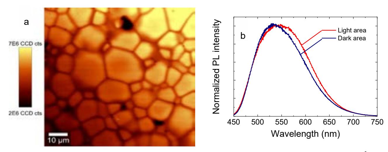
Our investigation revealed that GYAGG:Ce glass-ceramics samples all have a well-defined structure with clear boundries between crystallites (fig. 1). We also observed that between the aforementioned crystallites areas of reduced luminescence were present. These areas were roughly equivalent in size to the smallest observed crystallites and the luminescence measured from within those areas was less intense by a factor of 2. Spectroscopic measurements revealed that the shape of the photoluminescence spectra in the light and dark areas was similar, however the peak and the long-wavelength edge of the dark area spectra are blue-shifted by approximately 20 nm.
Similar distributions of photoluminescence parameters were observed in all samples. The density and shapes of the crystallites and dark spots are different. Density of dark spots increases with increased sample density. The results of this study will be usefull for determination of optimal production technology for GYGAGG:Ce glass-ceramics