
Single-walled carbon nanotubes (SWCNTs) are among the most promising nanomaterials for theranostics. Due to their specific physico-chemical characteristics SWCNTs have the potential to be used both for cancer cell imaging [1] and destruction [2]. Carbon nanotubes are supposed to be used in bioapplications as photoacoustic imaging contrast agents or as photothermal therapy heating agents [3]. Under the influence of pulsed laser radiation on an absorbing medium, its local heating, thermal expansion, and generation of compression and rarefaction waves occur.
In present study we show the effect of continuous and pulsed laser irradiation of near-infrared (NIR) spectral range on rat C6 glioma cells in three different model systems: 1) cells, accumulated SWCNTs inside the cytoplasm as small agglomerates of μm size, 2) cells, exposed to nanofluid of separately dispersed single (or small bundles of several nanotubes) SWCNTs in non-covalent complexes with DNA molecules, 3) cells without SWCNTs either inside or outside the cell.
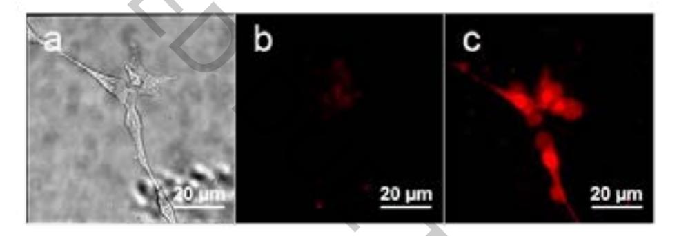
By means of Raman spectroscopy we demonstrate the accumulation of SWCNTs inside the cell cytoplasm in the form of μm-sized agglomerates presumably in the endoplasmic reticulum after 24 h of cells exposure to SWCNTs. Glioma cells irradiation with lasers of $\lambda_{ex}$ = 635 nm and $\lambda_{ex}$ = 785 nm in continuous mode does not influence cell viability neither in the presence of SWCNTs nor in their absence, excluding the probability of cell destruction due only to the conversion of absorbed radiation by carbon nanotubes into heat. Cell viability was detected by fluorescence microscopy using. Exposure of glioma cells with SWCNTs accumulated inside the cell to pulsed irradiation with $\lambda_{ex}$ = 909 nm and picosecond laser pulse duration leads to effective cancer cell destruction (Fig. 1), while similar impact on cells in a medium with dispersed individual nanotubes does not affect their viability. We propose theoretical model of vis-NIR laser irradiation interaction with SWCNTs and cancer cells. We consider the problems of energy absorption of pulsed laser radiation by cylindrical and spherical nano- and microparticles (SWCNT individually and in agglomerates, respectively), which differ in physical characteristics from the environment (biostructure). The results of simulation confirm that photo-induced destruction of cancer cells with picosecond pulsed irradiation in NIR spectral range can be achieved only when SWCNTs are accumulated inside the cell, but not when SWCNTs are dispersed in the external medium (under the same exposure conditions).