
Protein aggregation into amyloid fibrils is associated with several neurodegenerative disorders, such as Alzheimer's, Parkinson's, or prion diseases [1]. Amyloid aggregates are formed by conformational changes in the native protein structure and subsequent elongation [2]. Amyloidophilic dye molecules, such as thioflavin-T (ThT) and Congo red (CR), which bind to grooves formed by beta-sheets on the fibril's surface or 8-anilinonaphthalene-1-sulfonic acid (ANS) – which binds to the fibril's hydrophobic regions, can be applied to track amyloid formation and in some cases, such as methylene blue, they can act as potential aggregation inhibitors [3-5]. The changes to dye fluorescence intensity or absorbance spectra are considered to be caused by an increase or decrease in the concentration of fibrils. However, this could be the result of how such molecules interact with the fibril's surface or with one another, leading to inaccuracies in amyloid assays. In this work, the interaction of amyloidophilic molecules on the surface of amyloid fibrils was investigated using absorption and fluorescence spectroscopy.
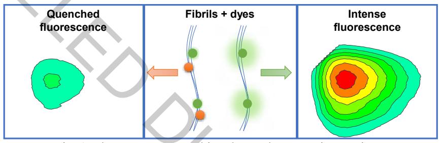
Insulin, lysozyme and mouse prion protein amyloid fibrils were prepared at 60°C. The absorbance spectra and excitation-emission matrices (EEM) of the samples were measured after mixing the fibrils, dye and PBS in the range from 200 to 800 nm. In many cases, amyloidophilic molecules, such as ThT, CR, Dapoxyl, ANS or MB assist each other in binding to amyloid fibrils, but this does not increase the fluorescence intensity of ThT, ANS, Dap. Often, there is a noticeable decrease in fluorescence (Fig. 1). Similar effect was observed using all three proteins.