
Carbon nanotubes (CNTs) are promising systems for biomedical imaging, diagnostics, therapy and can be applied as a component of biosensors and implants. Thereby, it is necessary to study the effects of CNTs on various biological systems and such investigations underway in various research centres [1–5]. The different types of polymer molecules can be used to increase the dispersibility of CNT suspensions and other purposes. The aim of this work is to identify changes in the structure and properties of human erythrocytes exposed to CNTs functionalized by polymers.
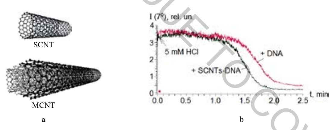
Carboxylated multi-walled CNTs (MCNTs) with a length of about 1 μm and single-walled CNTs (SCNTs) with a length of about 400 nm synthesized by the CVD method were used (fig. 1, a). Polyethylene glycol (PEG, unionogenic hydrophilic polymer) and deoxyribonucleic acid (DNA, anionic polymer) were non-covalently attached to CNTs. Erythrocytes were isolated from healthy donor blood by washing in 0.15 M NaCl and suspended in Earl's balanced saline solution (pH 7.3). MCNTs-PEG or SCNTs-DNA were added into erythrocyte suspensions up to the final concentrations from 1 to 100 μg/ml and incubated at 37 °C. The geometric parameters of erythrocytes after treatment with CNTs were studied by nephelometry and light microscopy methods. The levels and type of hemoglobin in extracellular medium of erythrocytes were determined by analysis of absorption spectra at 380–700 nm. The kinetic dependencies of HCl-induced erythrocyte lysis were measured as the kinetic dependencies of light scattering intensity at an angle of 7° in order to evaluate deformability changes of the cells exposed to MCNTs-PEG or SCNTs-DNA.
Significant impact of MCNTs-PEG or SCNTs-DNA at concentration of 1 and 2.5 μg/ml on erythrocytes were not revealed. It was found that MCNTs-PEG and SCNTs-DNA at high concentrations of 5–100 μg/ml induce deformation and damage of erythrocytes and an increase of hemoglobin release into extracellular medium from cells. These changes were more significant at the highest concentration of CNTs. The partial transformation of oxy-form of hemoglobin to met-form has been detected at action of MCNTs-PEG but not at influence of SCNTs-DNA. The acceleration of acid lysis of erythrocytes was observed after preincubation of the cells with SCNTs-DNA at high concentrations (fig.1, b).
The obtained data indicate that SCNT-DNA and MCNT-PEG can have a cytotoxic effect on human erythrocytes. The cytotoxicity of CNTs against erythrocytes is also observed by other researchers [2–5]. Moreover, interaction of CNTs with cells is depends on their length and physical and chemical surface properties as is found in our research particularly.