
Metal nanoparticles with different sizes and shapes have broad spectrum of application including localized surface plasmon resonance (LSPR), surface enhanced Raman scattering (SERS) [1], sensors, optical probes, catalysts [2]. The polyol synthesis route allows to obtain nanoparticles of various shapes and sizes. Size and shape of the nanoparticles mainly depends on the synthesis temperature, precursors, synthesis time [2,3].
Polyol synthesis is simple chemical reduction method where the medium of reaction is multivalent alcohols [4]. The main advantage of this synthesis is that variety of polyols can be used and simplicity - it does not require very expensive materials to obtain particles of normative size by chemical reduction in a short time.
In this research, polyol synthesis was used to synthesize silver nanostructures. The precursor was silver nitrate, copper chloride was used as etchant and polyvinylpyrrolidone as capping agent. All reagents were dissolved in 1,5-pentanediol. The reaction took place in a heated vial with 1,5-pentanediol repeatedly injecting AgNO3 and PVP solutions. These two solutions were injected repeatedly in a hot flask with 1,5-pentanediol. The process took about 15 minutes in average until the solution in reaction flask became opaque. The final solution was left to cool down in a room temperature and optical properties were measured (Avantes, 190-1100 nm, resolution 1.4 nm). The influence of synthesis temperature to the yield and geometry of nanoparticles was investigated in a range of 125 – 155°C.
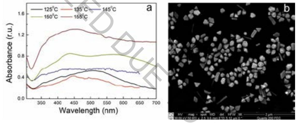
The effect of the temperature on the optical properties of colloidal solution is shown in Fig. 1 (a). It was found that in all used temperature range, silver nanostructures were formed as characteristic surface plasmon peak was observed. Only at 145°C temperature the peak is not so well expressed (due to broad size distribution). From the absorption spectra of the nanoparticles synthesized at 125 °C and 150 °C two main peaks were identified – at the 420 nm and 510 – 600 nm with shoulder at 350 nm. That is an indication of synthesized particles with sharp edges [5]. Analysis with SEM has proved our conclusion from UV-VIS spectra (Fig. 1 (b)). From the SEM image one can see that there are different shape particles and some of those are single crystalline particles and some – multiply twinned.