
Photosensitized tumor therapy is one of the most perspective methods to treat and diagnose oncological diseases. With the help of tetrapyrrolic compounds as photosensitizers and their retention of tumor tissues, it is possible to diagnose cancer in the early stages. In many cases of oncological diseases, this is the most important condition to successfully treat it. Photosensitizer is excited by the light after it accumulates in the tumor tissue and then can be used to localize cancer through the process of fluorescence. Moreover, singlet oxygen, which actively reacts with surrounding molecules that are in tumor tissues and damages them, is generated after the excitation of the tetrapyrrolic compounds [1].
Two tetrapyrrole compounds that initiate photooxidative reactions via singlet oxygen pathway were studied in this work: protoporphyrin IX (PPIX) and hematoporphyrin dimethyl ester (Hp-DME). A greenlight (532 nm) laser and a (405 nm) light emitting diode were chosen to excite the photosensitizers, the samples were dissolved in aqueous buffer solution (pH 7) with ascorbic acid and the spectroscopic data were registered using a EPR spectrometer “Bruker ELSYS E 580" and an absorption spectrometer “AvaSpec 2048".
Stock solutions were prepared with 10-3 – 10-4 mol/L concentration PPIX and Hp-DME. During the experiment, the acidity and concentration were varied in accordance with experimental needs.
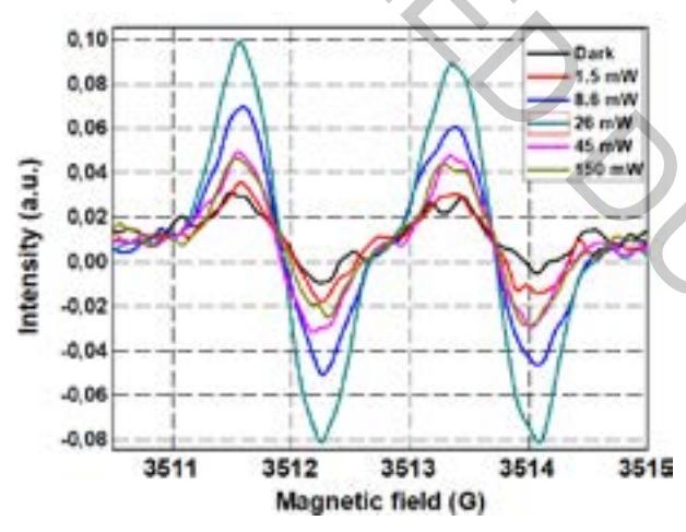
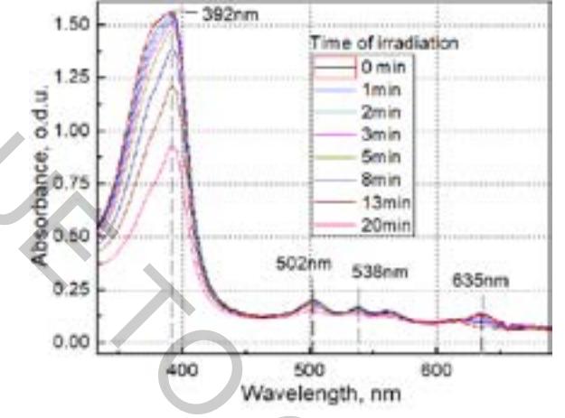
Nonlinear dependence of ascorbate radical generation on the intensity of laser radiation was registered during the examination of PPIX by applying EPR spectrometry. The amount of ascorbate radical increased while the intensity of the laser was increasing from 1.5 mW to 26 mW (Fig. 1). The registered amount of radicals started to decrease when the radiation intensity increased furthermore. Absorbance spectra of Hp-DME showed that the intensity of peaks decreased depending on the duration of irradiation (Fig. 2).
The relationship between EPR and absorbance spectra of PPIX and Hp-DME and their dependencies on acidity, concentration of antioxidants and, the intensity of various excitation sources will be presented and discussed.