
Metal chalcogenides are important because of their physical, chemical, optical properties and extensive potential applications in a lot of research areas [1]. Because of their different types of morphology, metal chalcogenides are applied for solar cells, Li-ion battery, Na-ion battery, photodetector, gas sensor and one of the newest application – ethanol gas sensor [1], [2], [3]. Tin compounds are abundant in nature and relatively cheap. For the fabrication is not needful expensive materials and toxic conditions.
The preparation of tin(II) sulfide was made using SILAR method. This method have some advantages, such as simplicity, easy to control the thickness and the composition of the films. For the synthesis of SnS films as a substrate FTO glass slide was used. Glass slides were ultrasonically cleaned in acetone at 40 °C, then washed with water and dried in air. For the SILAR cycles as a cationic precursor 0.1M tin(II) chloride acidic with a pH=1.5 (marked ac.) or alkaline solution with a pH=8 (marked al.) at 40 °C was used. As an anionic precursor 0.1M sodium sulfide solution at 40 °C was used. Firstly pre-cleaned substrate was immersed in the cationic precursor for 30s and tin ions adsorbed on the top of the substrate. Then sample was immersed in the anionic precursor for 30s, where sulfide ions react with adsorbed tin ions, and lastly in room temperature distilled water for 20s. 20 such deposition cycles were repeated in order to get adherent films. The last step was dip in the cationic precursor for 30s, then washed with distilled water and dried in air. The XRD technique was carried out by using a Bruker AXS D8 Advance diffractometer. EDX analysis was carried out using QUANTAX with X-Flash Detector 3001 and ESPRIT software.
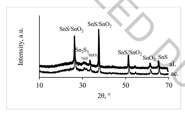
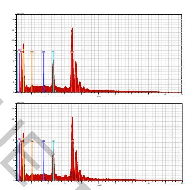
Figure 1 shows the results of X-Ray diffractions analysis and here it could be seen that the phase composition of both samples is very similar. The most intensive peak is at $2\theta$=37.8° and assigned to SnS (JCPDS card number 83-47). This peak also could be attributed to SnO2 (JCPDS card number 46-1088), which is on the glass slide. Other peaks with a similar situation are at $2\theta$=26.59 and 51.5°. There are two small peaks at $2\theta$=31.8 and 65.5°, assigned to SnS. Both samples have one peak at $2\theta$=30.8°, assigned to Sn2S3 (JCPDS card number 72-31). Based on the article [4], it is possible to get tin(III) sulfide at almost room temperature. From diffractograms it is clearly seen that using the cationic precursor alkaline solution the peaks of SnS are a little bit more intensive than by the use of acidic cationic solution.
Figure 2 shows the EDX results of both samples. There are two high peaks of tin and sulfur, which clearly show the formation of tin sulfides. Also, there is the peak of oxygen, showing the existence of tin oxide, which is on the top of the substrate. There also is a very small amount of sodium, left from the anionic precursor.
Based on XRD analysis it can be concluded that there is no strong difference between tin(II) chloride solution pH in order to get tin(II) sulfide films. The main products are the same, but the peaks of target products are a little bit higher using the alkaline solution of cationic precursor.