Amniotic fluid is a promising source of stem cells in regenerative medicine, since these cells are easy to isolate from amniocentesis samples, they display high proliferation potential as well as the ability to differentiate towards lineages from all three germ layers [1]. As neural tissue has restricted regeneration potential, amniotic fluid stem cells (AFSC) could be an attractive option for therapeutic purposes. Therefore the aim of this study was to evaluate the potential of AFSC to differentiate towards neural cell lineage under different induction conditions.
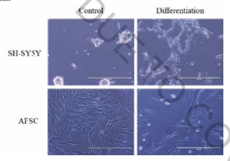
Stem cells were obtained using a two-stage isolation protocol and expanded in monolayer culture where they displayed typical spindle shaped morphology. Cells were characterized and were positive for pluripotent stem cell markers (Nanog, Sox2, Oct4, Rex1) and mesenchymal stem cell markers (CD44, CD73, CD90, CD105, CD146, CD166). In order to initiate neural differentiation of AFSC we used several induction protocols (Fig. 1) which comprised commercial supplement NeuroCultTM and biomolecules such as NGF, BDNF, cAMP, IBMX, retinoic acid and KCl. Later the expression of neural genes (NES, NSE, TUBB3, GFAP, etc.) was examined. Results of this study revealed some differences in expression of specific genes when using distinct combinations for neurogenic induction. In addition, the dependence of differentiation efficiency on chosen differentiation inducing agents was highlighted. Differentiated cells were also characterized by specific proteins (Tubulin B3, Vimentin and NCAM) using fluorescence microscopy. As a positive control for neuronal cell culture model human neuroblastoma SH-SY5Y cell line [2] was used.
The results of this work bring new insights about the ability of AFSC to differentiate into neural-like cells and the importance of differentiation induction conditions, however, a more in-depth research is required to acquire functional AFSC-derived neural cells.