
Luminescent semiconductor nanocrystals known as quantum dots (QDs) have widely been used in various biological assays: bioimaging, cell functioning, and intercellular interactions. [1] However, their 2D analogous – semiconductor nanoplatelets (NPls) - could be even more perspective in those applications. Having larger two-photon cross section comparing to both quantum dots and traditional organic fluorophores, they may be used as highly efficient fluorescent labels in biomedicine.
In frame of earlier established one order of magnitude larger two-photon absorption coefficient for CdSe nanoplatelets as compared to quantum dots we examined their efficiency for cell labeling and visualization with two-photon fluorescence microscopy. In order to do that we synthesized CdSSe/ZnCdS core-shell nanoplatelets and CdSe/ZnS quantum dots, both emitting at 585 nm. Those nanoparticles were then encapsulated with amphiphilic polymeric shell, having on their surface zwitter-ionic pairs and slightly positive zeta potential. That value is required for better penetration of charged nanoparticles in cells. [2] Glioma C6 cells incubated either with nanoplatelets or quantum dots demonstrated nearly equal uptake efficiency in the flow cytometry despite of sufficiently different dimensions of these two types of nanoparticles. Two-photon confocal fluorescence microscopy revealed ca. order of magnitude larger fluorescence response from nanoplatelets taking into account difference in concentration of nanoplatelets and quantum dots uptaken by cells.
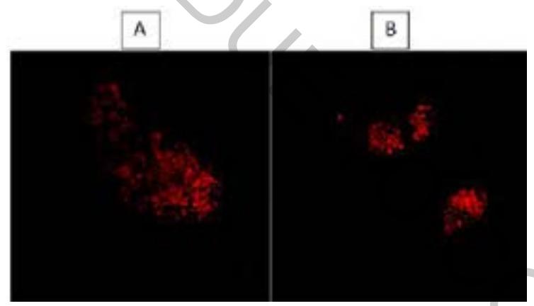
Our experiment confirms much higher labeling efficiency by nanoplatelets toward their visualization through two - photon fluorescence relatively to quantum dots which opens the door to use them as highly efficient fluorescent labels in biomedicine.