Nowadays, a variety of methods that can be applied for identification of different species of pathogenic fungi and bacteria are currently being sought and tested. Bacteria and fungi can cause infectious diseases, so it is important not only to identify them early but also to accurately determine their species. The early identification of fungi and bacteria in the organism can prevent a patient from negative consequences and it can help while prescribing antibiotic therapy, because antibiotics have no effect on fungi while some bacterial species can develop antibiotic resistance. Identification of fungi and bacteria is also important in food quality control, pharmaceutical and cosmetic manufacturing processes to ensure that the final product is not contaminated [1-3].
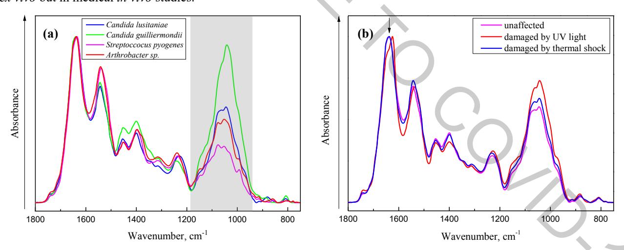
In this work, the method of an attenuated total reflection of infrared radiation (ATR IR) spectroscopy was applied for the analysis. ATR IR absorption spectra of 56 samples of different bacteria (20 samples, 9 different species) and fungi (36 samples, 5 different species) were analyzed. After spectral analysis, the main spectral differences that allow separating bacteria from fungi were observed in 1183-930 cm-1 spectral range (Fig. 1. (a)). The spectral bands which are observed in this range, varies depending on the fungi and bacteria species, and can be attributed to glycogen, amino acids and phosphates.
To assist the identification of bacteria and fungi from ATR IR spectra, cluster analysis was performed. Cluster analysis was performed using the Ward Algorithm, selecting the 1718-935 cm-1 spectral range and applying the first derivative and vector normalization preprocessing. Fungi and bacteria were separated into two clusters with 100% accuracy.
Fungi life-cycle analysis was carried out by performing cluster analysis, of spectra of normal, thermally damaged and damaged by UV light Candida lusitaniae fungi (Fig. 1. (b)). Cluster analysis was performed in 1725-1488 cm-1 spectral range using the Ward algorithm. Cluster analysis of C. lusitaniae showed that normal, damaged by UV light and thermal shock fungi can be separated with 100% accuracy.
In conclusion, it can be stated that infrared spectroscopy is a suitable method for the identification of bacteria and fungi. More detailed studies with a larger number of the samples would let to apply infrared spectroscopy not only in ex vivo but in medical in vivo studies.