
Silver nanoparticles (Ag NPs) have become attractive for various electrooptical applications because of their plasmonic properties. These properties originate from the excitation of oscillations of free surface electrons, a phenomenon in nanosized plasmonic metals known as Localized Surface Plasmon Resonance (LSPR) [1]. Metal NPs with unique optical properties can be created by varying size and shape of NPs therefore better control of these parameters could lead to more advanced applications [1].
The purpose of this work was to modify chemically synthesized Ag NPs using ultrafast laser pulses and analyze the dynamics of ultrafast relaxation processes after the modification. Ag NPs were synthesized employing polyol synthesis method using 1,5-pentanediol as reaction medium, silver nitrate AgNO3 as precursor and PVP as capping agent [2]. Synthesized nanoparticles had broad size distribution and cube like shape where the length of cube edge was ranging from few nanometers up to 60 nm. The application of ultrashort laser pulses was used in order to change the size distribution expecting it will become narrower.
The modification of colloidal solutions was performed with femtosecond Yb:KGW laser (290 fs pulse duration, 1030 nm wavelength, 40 kHz repetition rate, 4 W power) Steady-state absorption measurements were performed with an UV-VIS spectrometer (Avantes) in a range of 200 – 1000 nm (Fig. 1 a). Ultrafast transient absorption spectroscopy (TAS) technique was used to measure ultrafast plasmon relaxation dynamics in Ag NPs. The size distribution of Ag NPs was determined from SEM micrographs using “ImageJ" software (Fig. 1 b, c).
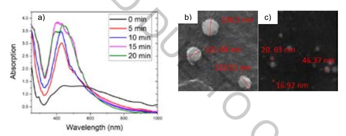
The Ag NPs affected with femtosecond laser pulses for 0, 5, 10, 15 and 20 min were analyzed by TAS, UV/VIS steady-state absorption spectrometer and SEM. TAS spectra have not shown a clear dependence on laser irradiation time of Ag NPs samples. However, it has an influence on decay times of TAS signal relaxation. The deeper analysis of absorption graphs (Fig. 1 a) and SEM micrographs (Fig. 1 b and c) revealed that the absorption spectrum of the laser affected Ag NPs was shifted into shorter wavelengths area and the peak width was reduced in comparison to primary Ag NPs sample. The irradiation with ultrashort laser pulses resulted in the change of nanoparticle shape into spherical. Depending on the irradiation time average Ag NPs diameter varies from 130.38 nm (5 min) to 40.64 nm (20 min).