Urinary bladder cancer is the 10th most diagnosed form of cancer in Lithuania and worldwide. This type of cancer affects about 4 times more men than women. By 2030, the number of new bladder cancer cases in Lithuania is expected to increase by 16 % for men and 62 % for women [1]. Cystoscopy (bladder endoscopy) with the following biopsy of the suspicious areas of the bladder and cytological examination of the bladder washouts are the most commonly used methods in the diagnosis of bladder cancer [2]. Histological examination of bladder tissue today is the golden standard for diagnosing bladder cancer but it is an invasive, time consuming procedure. Furthermore, cytological examination is effective for the diagnosis of tumors with a high degree of differentiation, but have low susceptibility to the most common tumors of low malignancy and early stage. As a result, bladder surgery makes it difficult to determine the exact limits of tumor expansion intraoperatively. Effective identification of bladder and other cancers of the urinary system and the establishment of clear boundaries between healthy and tumor tissue require new reliable diagnostic techniques that enable rapid and accurate characterization of samples.
Tumor growth and metastasis lead to changes in the concentration of certain molecules in the body. These changes can be monitored by quantitative and qualitative analysis of biological fluids. One such fluid whose changes in composition are associated with metabolism of cancer is the extracellular fluid [3]. It involves the supply of nutrients essential for cellular metabolism and the removal of metabolites from them. The extracellular fluid layers are formed by smearing the tissue sample across the specimen holder. Single tissue cells may also be found in the layer of extracellular fluid formed in this way. For this reason, extracellular fluid assays provide information not only on the changes in the metabolism of the tissue cells, but also on the morphological differences between the cells themselves.
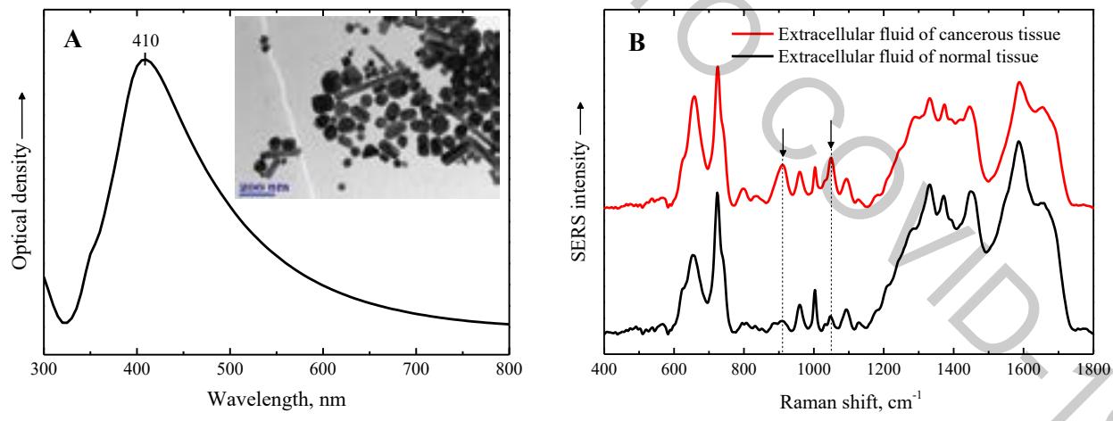
Molecular vibrational spectroscopy techniques are well-suited for detecting changes in molecule concentration which are caused by cancer activity. The aim of this work was to investigate the applicability of the surface enhanced Raman scattering (SERS) spectroscopy for the detection of cancerous urinary bladder tissue. By using this non-destructive method, the Raman scattering signal is enhanced when the analyte molecules approach the metal-dielectric phase boundary or adsorb on the metal surface. Colloidal solutions of noble metal nanoparticles are commonly used to form SERS active surfaces (Fig. 1 A). The effectiveness of these layers depends on the size, shape, concentration, type of the metal and other factors. Optimization of the synthesis of colloidal solution is an important process that can improve the quality of SERS research. Thus, the application of SERS spectroscopy for the analysis of biological tissue and extracellular fluid could have a direct application in the diagnosis of oncological diseases (Fig. 1 B).