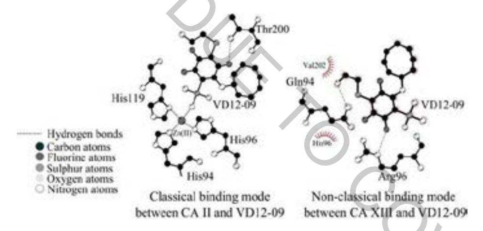
The mode of sulfonamide binding to human carbonic anhydrases has been demonstrated in more than 500 crystallographic structures deposited in PDB database [1]. This binding mode is characterized by the formation of coordination bond between sulfonamide nitrogen atom and Zn(II) cation located in the ~15 Å-deep cone-shaped cavity formed of a hydrophobic and hydrophilic regions [2]. However, we were able to obtain several unusual crystal structures of CA XIII in complex with ortho-, para- disubstituted fluorinated primary sulfonamide compounds (VD12-15, VD12-09) where the fully reversed orientation of the inhibitor was observed. There was no coordination bond and the sulfonamide group was exposed towards the solvent.
Several CA mutants were generated utilizing PCR-mediated site-specific mutagenesis in order to determide structural reasons for such unusual ligand positioning. Two recombinant active site mutant proteins (CA II Thr199Val, Ca XIII Val201Thr) were constructed in this study. For expression of the active site mutant proteins, plasmids carrying mutations were transformed into E.coli. proteins were purified by IMAC, ion exchange and affinity chromatography. Fluorescent thermal shift assay (FTSA) was used for evaluation of dissociation constants. Three X-ray crystallographic structures of mutated protein and ligands were solved and analyzed.
Despite the hypothesis, we were unable to detect reverse-orientation ligand positioning in the active site of mutated proteins. Consequently, we decided that unexpected ligand position was an artifact due to acidic pH (4.0) buffer used for crystallization. However, mutation in CA II active site did not influence ligand upside down positioning towards solvent. Still, one amino acid substitution in protein active site could influence the binding affinities in mutant proteins in some cases.