Heterogeneity is an intrinsic property of cell's structure and most biomechanical parameters. The key of such parameters – cellular viscosity, maintains cellular structure, regulates diffusion of biomolecules and significantly alters cellular behavior. Various cell mechanisms, including the differentiation of mesenchymal stem cells (MSC), apoptosis or diseases, e.g. cancer, diabetes or Alzheimer's disease results in the change of viscosity within plasma membrane and other organelles [1]. The ability to measure and visualize viscosity gradients would provide greater insight into the vital processes or even allow us to achieve earlier diagnosis of different diseases. Although there are few fluorescence-based methods for viscosity determination at the cellular level, e.g. fluorescence recovery after photobleaching (FRAP) or fluorescence correlation spectroscopy (FCS) – utilizing them would only result in single-point measurements of viscosity, which is insufficient for creating intracellular viscosity 'maps'. Therefore, a novel method is required for a spatial resolved quantitative viscosity ‘mapping' in biological objects.
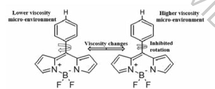
Viscosity-sensitive fluorophores ('molecular rotors') are the benchmark for viscosity bioimaging. One of such molecules - BODIPY-h is based on BODIPY (4,4-difluoro-4-bora-3a,4a-diaza-s-indacene) fluorescent dye. Phenyl ring's rotation in the BODIPY-h molecule depends on the viscosity of the surrounding micro-environment (Fig. 1): in a less viscous medium, higher intramolecular rotation gives rise to quicker deactivation from the fluorescent state via non-radiative pathway, thus resulting in a shorter fluorescence lifetime and vice versa [1], [2].
The aim of our study was to determine absorption, fluorescence spectra and fluorescence lifetime of BODIPY-h in cell growth media - Dulbecco's Modified Eagle Medium (DMEM) with fetal bovine serum (FBS) and phosphate-buffered saline (PBS), together with the uptake of the molecular rotor in human skin mesenchymal stem cells and their differentiated counterparts: adipocytes, osteocytes and chondrocytes.
Absorption, fluorescence spectra and fluorescence lifetimes of BODIPY-h were measured in PBS and cell growth media - DMEM with FBS. Fluorescence lifetimes were also measured in human skin MSC suspension with Edinburgh FLS 920 fluorimeter. For the uptake evaluation, StemPro differentiation kits (Gibco, US), were applied for specific differentiation of MSC. Cells were stained with 9 μM BODIPY-h solution diluted with DMEM (1:1000) (Gibco, US) and incubated for 60 min. The accumulation of dye was observed using Nikon Eclipse Te2000-U, confocal microscope (Nikon, Japan).
Results revealed that BODIPY-h interacts with proteins - shifting its absorption and fluorescence spectra by 5 nm and 10 nm, respectively. Fluorescence lifetime kinetics showed the same interaction: fluorescence lifetime in medium without FBS is monoexponential, while with protein it becomes biexponential and grows from 212 ps to almost 4 ns. Uptake evaluation revealed a difference in BODIPY-h localisation between undifferentiated MSC and already differentiated adipocytes. BODIPY-h accumulated in the lipid droplets, formed within the adipocytes. Meanwhile, in osteocytes, chondrocytes and undifferentiated MSC, localisation of the dye was different – molecular rotor diffuses through the membrane, stains membrane-bound organelles but is unable to pass the nuclear membrane.