Pancreatic cancer has one of the highest mortality rates [1]. Pancreatic tumors tend to spread. Usually it spreads to liver, also it can metastasize to lymph nodes, lungs or other more distal organs. If a tumor is resectable, the standard treatment applied for pancreatic tumors is a surgery followed by chemotherapy. During the surgery, one of the most important factors for successful treatment is determination of the exact margin between normal and tumorous tissues. In order to avoid recurrence of the disease, all of the malignant cells have to be removed. On the other hand, it is important to leave as much as possible of the healthy tissue to safe vital functions of the organ. A method for real time identification of a tumor, usable directly during the surgery is needed.
During the last decades, various methods of vibrational spectroscopy were applied for biological tissue examination [2]. In case of conventional spectroscopic experiment, the sample has to be transferred to the device, the examination of tissue is limited by size and preparation of the sample. All these factors restrict the use of the methods for clinical application. Using the technique of attenuated total reflection of infrared radiation (ATR IR), the sample can be examined in native conditions without any special sample preparation. The use of optical fibers enables to perform measurements in situ or in vivo conditions, this is important during live surgery.
In this study, we used ATR fiber probe coupled with portable standard FT-IR spectrometer. The compact size of the device allows to perform the measurements in the operating room. Spectra of the tissue can be collected in a few minutes.
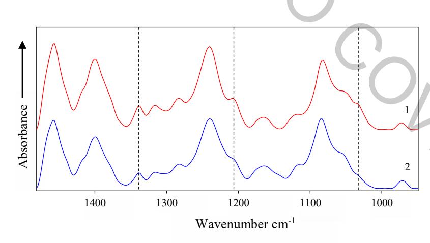
During the study, cases of 25 patients with pancreas pathology including pancreatic ductal adenocarcinoma, neuroendocrine carcinoma, cystic, metastatic tumors and pancreatitis were investigated. ATR IR absorption spectra of normal and tumorous (pancreatic ductal adenocarcinoma - PDAC) tissue are presented in Fig. 1. The most significant differences between normal and cancerous tissues are observed for the bands at 1034,1207 and 1339 cm-1. The increased intensity of these spectral bands in tumorous tissue indicates altered levels of collagen in the tissue. The amount of collagen in PDAC tissue increases due to the desmoplasia which is a common feature of PDAC tumors. During this desmoplastic reaction fibroblast cells secrete more fibrous proteins (collagen) in the extracellular matrix of the tissue. Established spectral markers for tumorous pancreatic tissue indicates that applied technique is appropriate for malignant pancreatic tissue identification.