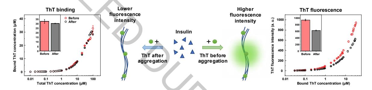
Amyloidogenic protein aggregation into fibrils is linked to several neurodegenerative disorders, such as Alzheimer's or Parkinson's disease [1]. An amyloid specific fluorescent dye thioflavin-T (ThT) is often used to track the formation of these fibrils in vitro [2]. Despite its wide application, it is still unknown how many types of ThT binding modes to amyloids exist, with multiple studies indicating varying numbers [3,4]. In this work we examine the binding of ThT to insulin fibrils generated at pH 2.4 and reveal a possible inner core binding mode which is not accessible to the dye molecule after aggregation occurs.
Insulin fibrils were prepared by incubating 100 or 200 μM insulin solutions (pH 2.4, 100 mM NaCl, 100 mM phosphate buffer) with or without additional ThT at 60°C without agitation for 24 hours. For each ThT concentration, excitation-emission matrices were scanned and used to determine both the maximum ThT fluorescence intensity and the position of the highest intensity peak. Absorbance measurements were used to determine the amount of free and bound ThT present in solution after sample centrifugation.
Insulin fibrils formed with ThT added before aggregation display an additional ThT binding mode, which is not accessible to ThT molecules after the fibrils are fully formed. ThT bound in this mode possesses a much higher quantum yield (Fig. 1) when compared to other bound dye molecules.