
Polydimethylsiloxane (PDMS) is one of the most widely used flexible polymers for various microdevices due to its physical flexibility, ease of processing, high chemical resistance, and high transmission in visible spectral region. Ultra-short femtosecond laser pulses have been shown to modify the refractive index of PDMS or even form microcavities when focused in bulk of material [1]. The formation of microcavities allows to form 3D structures in the volume of PDMS. This could be used to create 3D microchannels or optical elements inside PDMS without affecting its surface or using other, more complicated and time-consuming methods like soft lithography.
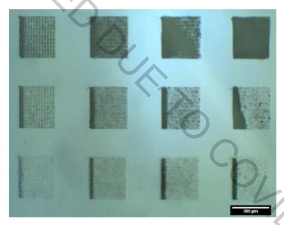
By changing pulse energy, pulse density and pulse repetition rate of the Yb:KGW femtosecond laser, we can choose between modifying PDMS refractive index or creating microcavities. Figure 1. depicts how the volume of the 3D cubes can be micromachined depending on the laser writing parameters, where cube in the bottom left corner, has the lowest pulse energy of $0.75 \mu J$ and pulse density of 50 pulses per mm, while cube in the top right corner has the highest, i.e. $3.75 \mu J$ and 250 pulses per mm, respectively. Three types of laser damages were identified from the bright field images of the optical microscope.
X-ray microtomography was employed for the characterization of 3D microstructures. It is a non-destructive method for investigation of local density changes in the volume of materials with micrometric spatial resolution.