A complete understanding of preimplantation stages of mouse embryo development is still lacking. A technical mainstay of research into the specific function of individual genes/proteins during these stages has been the use of antibodies to detect expression and sub-cellular localisation using microscopic immuno-fluorescent staining approaches [1]. However, this method is not 100% reliable, as antibodies are not always as specific as claimed and not all proteins have available and reliable anti-sera.
Hence, one option to study specific proteins is to create transgenic embryos that incorporate genetically encoded fluorescence reporter proteins as fusions with endogenous candidate genes [2]. Thus, permitting the expression level and subcellular localisation of specific genes to be assayed, across preimplantation developmental time, in both live and fixed embryos. We are currently adopting one such method to achieve this aim that exploits the CRISPR-Cas9 system to fluorescently tag candidate cell-fate related genes in mouse embryos.
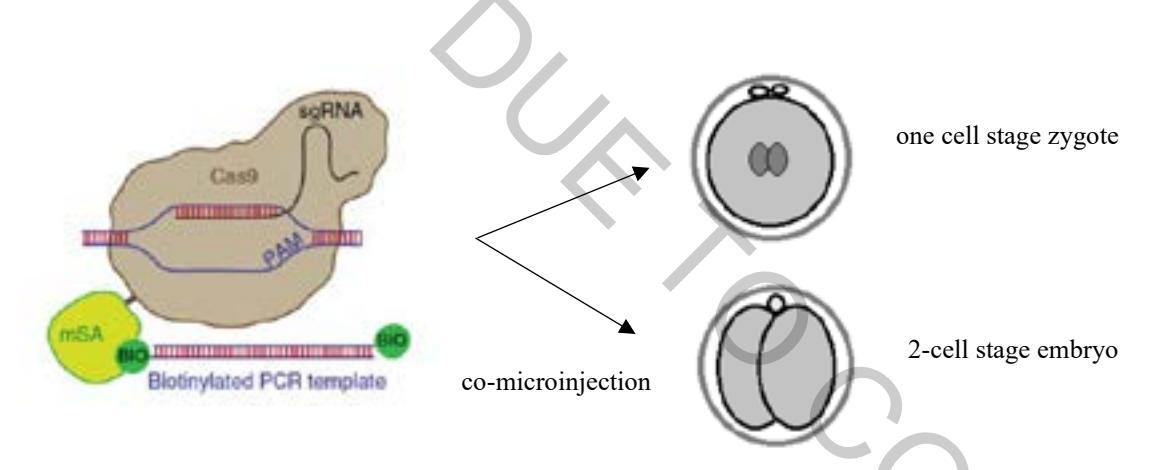
The main goal of my study is to create the necessary in vitro derived RNAs (i.e. biotinylated candidate gene specific guide RNAs/sgRNA and Cas9-Strepavidin mRNA) needed (Figure 1). Such RNAs will be co-microinjected into one cell stage zygotes or 2-cell stage mouse embryos, together with generated (by myself) and engineered recombinant DNA repair templates that target the candidate gene loci to ensure C-terminal tagging with the fluorescent protein mCherry. We will then microscopically assay the expression and sub-cellular localisation of these candidate genes, via the fused mCherry fluorescent tag, up to the peri-implantation blastocyst stages.