The ability to create silver nanoparticles with precise control of their physical, chemical and structural properties is crucial for expanding their applicability in various applications, e.g. optoelectronics, energy storage, catalysis [1], chemical and biological sensors [2], biomedicine [3] and others. Because of its properties, silver can be used in surface-enhanced Raman scattering (SERS) sensors, which allow the detection of vanishingly small amounts of various materials and, in some cases, single molecules [4-5].
The aim of this work was to synthesize spherical silver nanoparticles employing simple and reproducible chemical reduction method. Silver nitrate (AgNO3), tannic acid (C76H52O46) and trisodium citrate (Na3C6H5O7) were used as precursor, redactor and stabilizer respectively. The results of UV-Vis absorption spectra were recorded on an AvaSpec-2048 fiber optic spectrometer (172 - 1100 nm, 1.4 nm resolution). The size and shape of silver nanoparticles were determined by microscopic analysis using scanning electron microscope QUANTA 200FEG (1.2 nm (30 kV, SE)). The SEM images were analysed with ImageJ software [6] and size distribution of particles was determined measuring at least 50 particles in each image. The absorption spectrum of silver nanoparticles was theoretically calculated using the MiePlot program [7].
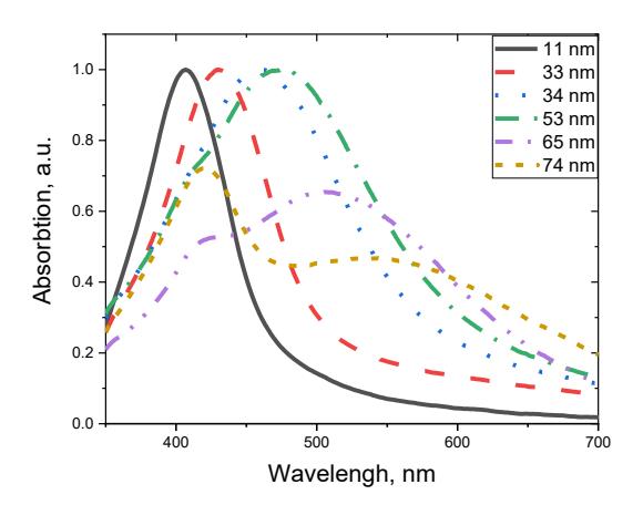
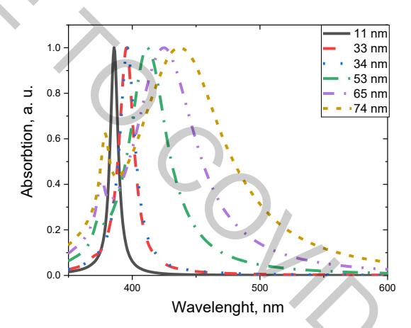
Colloidal solutions with small particles (11 nm diameter) appear yellow and clear and this solution was used as seeds for further growth of nanoparticles. After the growth process solutions with larger particles lose characteristic yellow colour due to the multipole plasmon resonance, which becomes the dominant spectral component. The UV-VIS absorption spectra of different size silver nanoparticles is presented in Fig 1. From the absorption spectra, one can see that the peak position is shifting towards longer wavelengths and becoming broader, suggesting that the particle size is increasing. When the size of Ag nanoparticles reach 53 nm, the absorption peak begins to develop a "hump" at 405 nm, which represents the quadrupole resonance of silver nanoparticles. SEM analysis has shown that synthesized particles prevail spherical shape and Mie scattering theory was used to model the optical response of the particles (Fig. 2). Comparing the theoretical and experimental results one can see that theoretically generated curves are much narrower compared to the experimental ones. This can be explained by the fact that in theoretical calculations only single value of particle radius was used and the experimental spectrum is a sum of signals generated of different size particles.