
Raman scattering spectroscopy is widely used for different applications that include chemistry, material science, medicine and biomedical sensing [1]. Since the discovery of the surface-enhanced Raman scattering (SERS) effect scientists have started to produce specialized substrates suitable for detection and study of extremely low concentrations of biomolecules by SERS spectroscopy. Most commonly, SERS-active substrates incorporate nanoscale noble metal particles of various shapes in colloidal solutions or placed on glass and silicon substrates. Certain plasmonic nanoparticle arrangements on the diffraction gratings have demonstrated an increase in the Raman signal enhancement up to a four-time [2].
In this work, we demonstrate a highly efficient combination of silver nanoparticles (AgNPs), porous silicon (PS) and diffraction grating for SERS application. Diffraction grating of 6-15 μm pitch was produced in n-type silicon wafer substrates by Yb:KGW femtosecond laser ablation. Different quality periodical lines in silicon where achieved varying the pulse densities and energies per pulse. Afterwards, the diffraction gratings were porosified by the electrochemical etching in an electrolyte based on a hydrofluoric acid. Silver colloids were synthesized following the Turkevich protocol [3] and exhibited a localized surface plasmon resonance related absorbance band in the blue range of the visible spectrum. Ag NPs were adsorbed on top of the PS grooves by exposing them to the colloid solutions for 1 hour. The same process was carried out for 3 days for periodical grooves formed on monocrystalline silicon wafers.
Morphologies of the produced structures were studied by scanning electron microscopy. Dependence of the resulting grooves' width on the number of applied laser processing parameters was determined. Major differences of the AgNPs deposition density on PS and monocrystalline silicon diffraction gratings were observed. SERS activity was evaluated using 3D scanning confocal Raman spectroscopy with a 633 nm laser and using Rhodamine 6G (R6G) as the test analyte.
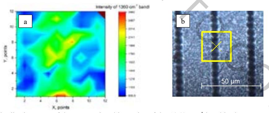
AgNP-covered PS diffraction gratings demonstrated expressed SERS signal of R6G. The enhanced of Raman scattering signal is two times higher in the grooves compared to the ridge of the investigated periodical structures (see Fig. 1). In contrary, pristine Ag NP-covered silicon diffraction grating was not demonstrating any useful signal enhancement.
In summary, new type of SERS substrates based on the AgNPs in diffraction gratings of porous silicon were demonstrated. The preliminary results of the enhancement of Raman scattering are promising towards development of methods for highly sensitive detection of organic compounds.