Bladder cancer is one of ten most frequently diagnosed cancer types worldwide [1]. Depending on tumor type and stage, conventional treatment of bladder cancer includes surgical operation combined with medication or radiation therapy [2]. The most frequent type of bladder cancer is urothelial carcinoma which requires surgical approach at the presentation in most of the cases. During the surgery, it is important to ensure the complete removal of the whole tumor in order to reduce the risk of recurrence of the disease. The gold standard method for tissue identification is histological examination of the removed tissue. The disadvantage of this method is that the specific sample preparation is required, also, this procedure is time consuming. Therefore, new diagnostic techniques are required. The intraoperative diagnostic technique should give fast and reliable results for precise determination of the tissue area which has to be removed and not to prolong the time of surgery to reduce the risk of complications.
During tumor formation, the metabolism of cells changes, therefore, it affects the chemical composition of the tissue. Vibrational spectroscopy is sensitive method for the qualitative and quantitative chemical analysis, thus chemical changes of the tissue should be observable in the vibrational spectra. In this study fiber based attenuated total reflection infrared (ATR IR) spectroscopy was applied for the tissue examination. Spectra could be measured within few minutes without almost any special sample preparation. Application of fiber probe enables to do measurements in situ or in vivo conditions, that is important during the surgery.
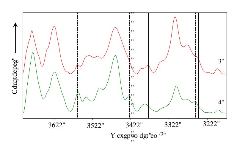
During the study, samples of normal and tumorous human bladder tissue of 49 patients were examined, including cases of urothelial carcinoma, nonspecific cystitis and papillary urothelial neoplasm of low malignant potential. The technique was applied for the tissues removed after the surgery. The ATR IR absorption spectra of normal and tumorous tissue are represented in Fig. 1. The decreased intensity of spectral bands of collagen located at 1339 cm-1, 1205 cm-1 and 1033 cm-1 is observed in the ATR IR absorption spectra of urothelial carcinoma tissue. It is related to the decreased amount of collagen in the tissue. The bladder is elastic organ that can dilate when the bladder is full and shrink when it is emptied, collagen provides the elasticity. During the process of tumor development, tissue loses its function, therefore, it could lead to decreased levels of collagen. Also, the increased intensity of the spectral bands located at 1155 cm-1 and 1026 cm-1 is observed, it is related to increased levels of glycogen. An increased amount of glycogen in the cytosol is a characteristic feature for some types of cancer. Glycogen is accumulated and used as the source of energy since cancerous cells due to high proliferation rate need more energy than normal ones.