Graphene oxide (GO) has the great potential for biomedical applications and environmental protection. The increased use of GO raises their release into the environment. However, the health risks associated with environmental exposure to GO are largely unknown. Due to their unique physiochemical properties, GO and its composites could be applied to wastewater treatment and to adsorptive remediation of environmental pollutants.
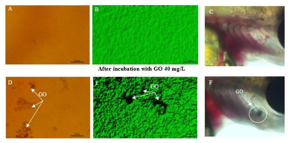
The aim of this study is to examine the distribution of GO 40 mg/L in Salmo trutta embryos and larvae using confocal fluorescence microscopy. Analysis was based on 3D autofluorescence imaging of test-organisms (Fig. 1. B and E). GO distribution in embryo chorion and external body tissues of larvae was evaluated. For the experiment, S. trutta embryos at the eyed-egg stage and 4 day old larvae were obtained from the hatchery (Lithuania). Studies have been carried out with non-protected life-stages accordance with EU Directive 2010/63/EU. The laboratory treatment was carried out in an environmental chamber (Bronson PGC-660, Zaltbommel, The Netherlands) with continuous aeration under static conditions.
The bright field image of control embryos show that the chorion surface is smooth with regularly distributed darker spots (Fig. 1. A). These darker spots are chorion pores that are important for oxygen/carbon dioxide, nutrients and excretion product transport to and from the embryo, respectively. After 4 days of incubation with GO 40 mg/L, in embryos chorion appeared non autofluorescence areas (dark spots), which indicated GO adhesion on surface of chorion (Fig. 1. E). Furthermore, dark spots of GO can be seen directly in some areas of embryos chorion and larvae internal tissues in the bright field image (Fig. 1. D and F). GO could clog the chorion pores and affect embryonic development of fish [1]. The question about non-homogenous GO adhesion on the organism is still open, because their biodistribution have not been fully investigated.