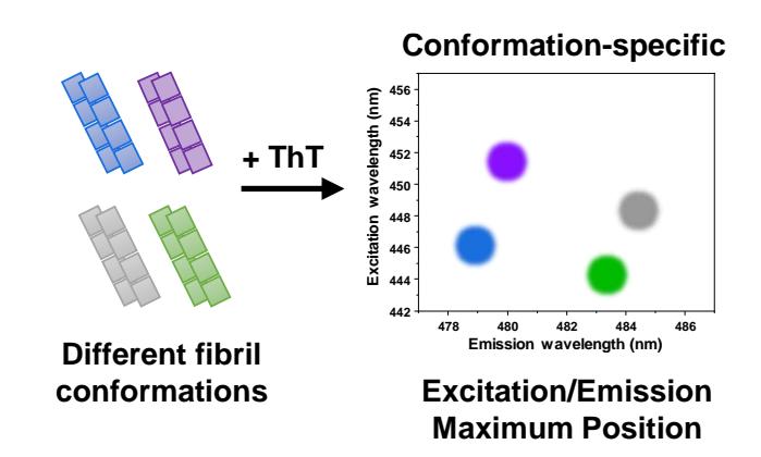
Amyloidogenic protein aggregation into insoluble fibrillar aggregates is linked to several amyloidoses, including neurodegenerative Alzheimer's and Parkinson's diseases [1]. Despite many years and countless studies, there are still very few effective anti-amyloid drugs available and most potential compounds fail to pass all clinical trials. One of the main reasons for these failures is the complex nature of amyloid aggregates. It is known that the same protein molecule may form fibrils with distinct conformational and morphological characteristics. Each of these fibril types also possess specific self-replication tendencies and may respond differently to potential anti-amyloid compounds. In many cases, fibril identification is done by employing atomic force microscopy, where results are highly dependent on sample deposition techniques, as well as infrared spectroscopy, which requires relatively high concentrations of the protein sample. In recent years it has been observed that distinct protein fibrils have specific amyloidophilic dye binding [2] characteristics (affinity, fluorescence intensity), which could potentially be used to identify unique amyloid aggregates.
In order to examine whether an amyloid-specific dye – thioflavin-T [3] could be used to differentiate between conformationally-different fibrils, insulin was aggregated into four distinct fibril types, under four environmental conditions. Human recombinant insulin powder was dissolved in four types of solutions (1. 20% acetic acid, containing 100 mM NaCl; 2. 100 mM sodium phosphate buffer (pH 2.0); 3. 100 mM sodium phosphate buffer (pH 2.4), containing 100 mM NaCl; 4. PBS (pH 7.4)) and incubated at 60°C. After fibrils were formed, they were initially examined by atomic force microscopy and Fourier-transform infrared spectroscopy, to verify morphological and structural differences. Afterwards, the samples were mixed with a range of thioflavin-T concentrations and their absorbance, as well as excitation-emission matrices were scanned.
The results show that each type of aggregate has unique bound-dye fluorescence properties, such as maximum excitation and emission wavelengths, as well as fluorescence quantum yield. Considering that such a dye-based examination requires minimal amounts of fibrils and is relatively quick to perform, it may be used as an alternative method to identify amyloid fibril conformation differences.