
Self-assembled monolayers (SAMs) of functional thiol molecules at a metal surface are widely used to study various functional groups' interactions with solution components or adjacent thiols in the monolayer [1]. Imidazole ring terminated molecules can thus interact with other aromatic residues as well as forming hydrogen bonds with polar and charged residues because they can exist in neutral or positively charged forms within a pH range of 5.0 to 7.0. The introduction of amide functionality in the hydrocarbon chain of adsorbing molecules greatly increases the stability of SAM due to hydrogen bonds forming between the adjacent molecular chains in the monolayer [2]. Interfaces between biologically active molecules and metals are an important issue in biocatalysis, biocompatibility, and biosensors. SAMs at metal surfaces provide a possibility to create stable molecular structures suitable for probing potential-driven molecular structure changes [3].
Surface-enhanced Raman spectroscopy (SERS) is a powerful spectroscopic technique to study molecular structure and interaction mechanisms between a terminal functional group of a monolayer and solution species at metal/electrolyte interfaces. Nonetheless, SERS is limited by SERS-active substrates (mostly Ag, Au, and Cu), and the requirement to use roughened or nanostructured surfaces restrict the applicability of this analysis method. Therefore, Tian et al. suggested a novel SERS technique named “shell-isolated nanoparticle-enhanced Raman spectroscopy” (SHINERS). The method is based on the enhancement of Raman signal by strong electromagnetic field provided by noble metal core nanoparticles surrounded by a few nanometer thick inert silica shell [4].
In this work, SHINERS was applied to determine the potential-induced changes in the molecular structure of N-(2-(1H-imidazol-4-yl)ethyl)-6-mercaptohexanamide (IMHA) at a smooth Au electrode. SHINERS method employing synthesized spherical silver nanoparticles with 85 ± 5 nm core size and SiO2 shell of 3 nm thickness.
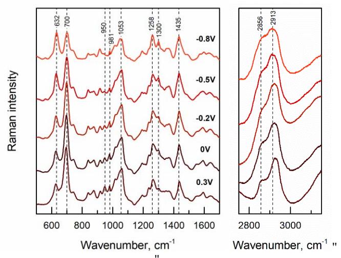
Spectroscopic evidence for potential-driven conformational changes in the structure of IMHA monolayer was revealed. In situ electrochemical SHINERS results show that the intensity of $v$(C-S)T mode (697 cm-1 at 0.0 V) of trans conformer is high at positive electrode potential value and noticeably decreases as electrode potential shifts to -0.8 V. The integrated intensity of the $v$(C-S)T band decreases by a factor of 2.3. The opposite of tendency with corresponding gauche band $v$(C-S)G (630 cm-1 at 0.0 V), the intensity of this mode increases by a factor of 1.46. These spectroscopic data indicate hydrocarbon chain conformational changes from predominant trans to gauche as electrode potential shifts to the more negative value.