
Recently bone grafts have been increasingly used around the world. Bone grafting frequency is indeed the second most frequent tissue transplantation worldwide, coming right after blood transfusion [1]. Over two million surgical surgeries are performed per year to treat bone tissue defects [1-2]. Bone tissue defects larger than 4-6 cm are critical and should be treated with bone grafts [2]. Critical-sized defects can be congenital or formed due to trauma, tumours, infections, and diseases such as osteoporosis, necrosis, or bone atrophy [1]. Four main types of bone grafting are used to treat bone tissue defects: autologous, allogeneic, xenogeneic, alloplastic. Autologous bone is currently considered the "gold" standard for treating bone tissue defects [1,3]. However, more and more attention is paid to alloplastic bone graft materials, as the use of these bone grafts could simplify operations and avoid the most common complications [1,3]. The major advantages of alloplastic materials include their high abundance relative to natural materials, no risk of disease transmission and the very low antigenicity. They can be made available in both resorbable and non-resorbable forms, and can be customized with varying levels of porosity and pore sizes [4].
The main objective of this study was to print hydroxyapatite (HA) scaffolds of complex geometry that could potentially be used to fill bone defects. One of the world's latest 3D ceramic printing technologies LMC (Lithography-based ceramic manufacture) was used for printing hydroxyapatite scaffold. Using this technology, the production of printed hydroxyapatite scaffold essentially consists of two main parts: 3D printing and heat treatment. The geometry of the printed HA experimental specimens (see Fig. 1), scaffold porosity, scaffold pore size, and material density fluctuations were analysed using industrial computed tomography (µCT). In order to analyse the structural properties of HA samples, studies were performed using X-ray diffraction (XRD) analysis and Fourier transform infrared spectroscopy (FT-IR) analysis. The surface topography and roughness of HA samples were analysed using optical profilometry.
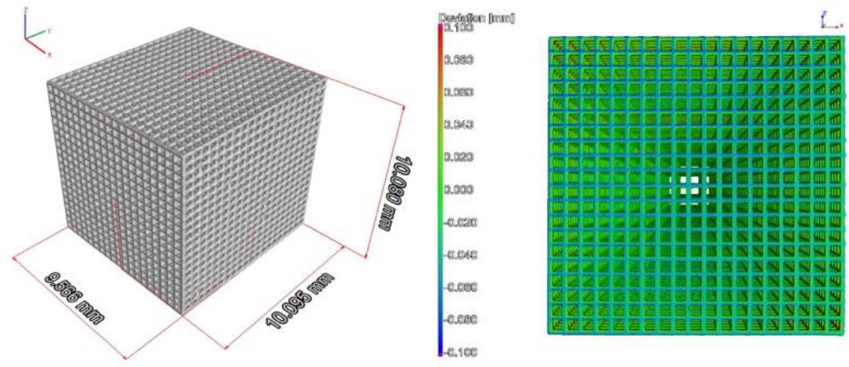
Based on the analysis of the results and comparison of the obtained results with the values found in the scientific literature as well as the requirements for bone grafts in medicine, it can be stated that printed HA scaffolds have a great potential to be used as synthetic bone grafts in the near future.