
Microbially induced calcite precipitation (MICP) for soil bioconsolidation is a relatively recently developed safe, effective and eco-friendly technique to improve the general properties of soils and solve several of the most common geoenvironmental problems, i.e. erosion, pollution and CO2 sequestration. This process utilizes the metabolic pathways of bacteria to form calcites (CaCO3) that bind soil particles together, leading to increased soil strength and stiffness because of this physical connection. The precipitation of carbonates via urea hydrolysis by ureolytic bacteria is the most straightforward and easily controlled mechanism of MICP with potential to produce large amounts of carbonates in a short period of time.
This study presents the analysis of Staphylococcus sp. H6 as a perspective biological tool to perform bioconsolidation of sand via MICP due to its high ureolytic activity. To our knowledge, we also present the first reported atomic force microscopy (AFM) analysis of an initial growth and precipitation of calcite on bacterial surface.
We conducted the analysis of Staphylococcus sp. H6 surface topography changes after treatment with cementation mixture using AFM. The obtained results indicated that the topology of Staphylococcus sp. H6 culture already changes after 1 hour's incubation with the cementation mixture (Fig. 1). It was observed that topographical changes begin forming from one pole of the cells (Fig. 1b, Fig. 1c, Fig. 1e), however this can be due to cell immobilization in the monolayer. It was hypothesized that amorphous CaCO3 precipitates were forming on the cells.
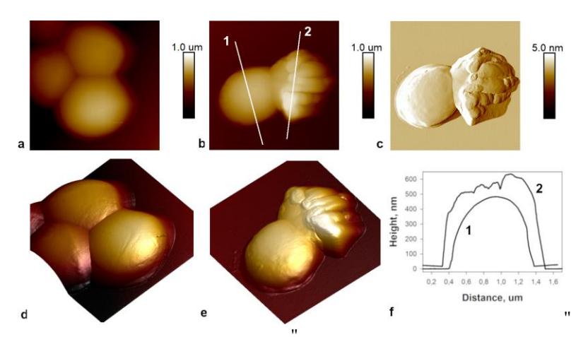
To test small-scale sand bioconsolidation, urease activity dependence on growth rate of Staphylococcus sp. H6 was first investigated. The small scale pilot studies of Staphylococcus sp. H6 application in bioconsolidation indicated that after exposure of sand with Staphylococcus sp. H6 culture solid sand column structures, or in some cases only platelets were formed. The structures obtained by using 0.5/1 M urea and 0.5/1 M CaCl2 displayed hardiness against H2O and can serve to clog pores in the soil and reduce soil permeability.
This report highlights a soil abundant Staphylococcus culture as a promising microorganism for MICP. Staphylococcus sp. H6 demonstrated attractive characteristics for CaCO3 precipitation and for sand particle bioconsolidation.