
Prostate cancer (PCa) is the most prevalent type of male cancer and the 5th leading cause of death in males with cancer worldwide in 2020 [1]. A large proportion of PCa is latent and does not tend to progress, whereas others are aggressive and significantly affect the life's quality. Therefore, it is of high importance to identify which PCa cases need immediate treatment while others could undergo active surveillance. Prostate-specific antigen (PSA) screening remains as a primary method for PCa diagnostics; however, it lacks specificity for other prostate diseases, such as benign prostate hyperplasia (BPH), and can often lead to false positive results and, in turn, unnecessary treatment. Moreover, it lacks prognostic value [2]. Abnormal changes in gene expression and epigenetic regulation are fundamental in any tumor type and lead to the development and further progression of PCa as well [3]. Altered histone methylation is one of the characteristic features during carcinogenesis, which may be due to altered expression of regulatory genes, like histone lysine methyltransferases (KMTs) and demethylases (KDMs). Therefore, histone methylation regulators might be utilized as effective biomarkers for more accurate diagnostics and characterization of PCa.
The aim of this study was to analyze the expression of KDM3A, KDM5D, KMT5A and PHF8 (a.k.a. KDM7B) genes associated with histone methylation in prostate tumors and control samples as potential biomarkers.
In the present study, 64 PCa, 25 noncancerous prostate tissues (NPT) and 16 BPH samples were analyzed for gene expression. The tissue samples were collected in 2008-2014 during the previous projects. First, total extracted RNA was used for the synthesis of complementary DNA. The latter was used for quantitative gene expression analysis by means of real-time PCR (qPCR).
The results of our study revealed differences in the expression of selected genes associated with histone methylation in PCa as compared to NPT and BPH. The expression of KDM3A in tumors and NPT was higher than that in BPH ($p = 0.0082$ and $p = 0.0164$, respectively), but it did not differ between PCa and NPT ($p > 0.0500$; Fig. 1A). KMT5A was highly expressed in BPH as compared to both PCa and NPT (both $p < 0.0001$; Fig. 1B), whereas PHF8 had generally low expression in all samples, nevertheless, it was significantly different between NPT and BPH ($p = 0.0206$; Fig. 1C). No associations were found between KDM3A, KDM5D, KMT5A and PHF8 expression and patient's age, prostate mass, serum PSA levels, tumor stage or biochemical disease recurrence (all $p > 0.0500$, not shown).
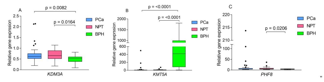
In conclusion, our analysis revealed significant differences in the expression of KMT5A, KDM3A and PHF8 genes in prostatic tissues, which suggest their potential utility for improved PCa diagnostics. However, their prognostic value was not determined. Additional investigation in larger independent cohorts is needed for further elucidation of the role of histone methylation regulatory genes in PCa.