
Polarimetric Second Harmonic Generation (SHG) microscopy can provide extra-molecular information from various non-centrosymmetric biological samples. The results of polarimetric SHG measurements require careful interpretation and modeling when the biological sample has a complex structure in the focal volume. In this study, a numerical modeling [1] has been performed to understand how biophysical parameters such as chiral (C) and achiral (R) susceptibility ratios are affected by different organizations of fibrillar structures in the focal volume. Then the results of numerical modeling have been used to interpret the images obtained from collagenous biological tissues and meso-tetra (4-sulfonatophenyl) porphin (TPPS4) molecules in giant “sea urchin” (GSU) aggregates using polarization-in polarization-out (PIPO) microscopy technique. The numerical modeling shows that in image plane crossing fibers [2] do not have a significant effect on the C ratio. The effect of crossed fibers on the measured R ratio is different for high and low molecular R ratio of individual fibers. In the case of low molecular R ratio, the effective R ratio increases with increasing the crossing angle between two fibers, while the effective R parameter decreases for high molecular R ratio crossing fibers.
On the other hand, tilting fibers out of the image plane affects both R and C ratios [3]. This geometry increases R and C ratios, and the C dependency has a sinusoidal behavior where the sign shows the direction of tilting. These results are in good agreement with the results obtained using PIPO measurements (Fig. 1).
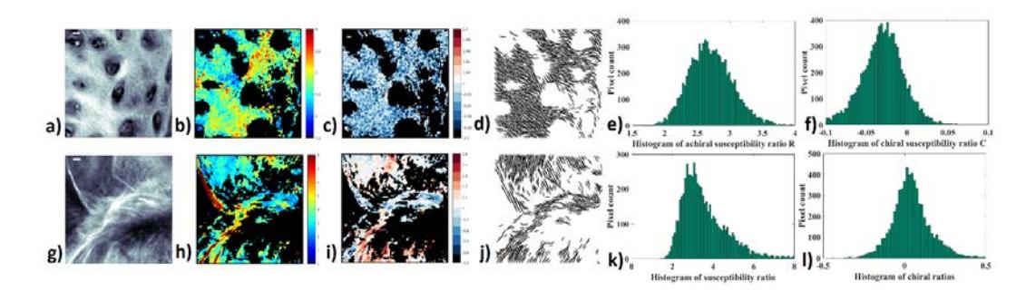
Fig. 1 a) and g) show the logarithmic scaled SHG intensity images of the collagenous and TPPS4 aggregate samples, respectively. Fig.1 b) and h) illustrate the results of R values calculated from Fig.1 a) and g). The TPPS4 has a higher R ratio than cartilage collagen. Fig. 1 c) and i) show the C ratios and Fig.1 d) and j) show the cylindrical orientation maps in the image plane. The histograms of R values are shown in Fig.1 e) and k). The histograms show a higher R ratio for TPPS4. The histograms of C values are shown in Fig.1 f) and l).
There are some regions of interest in the both samples with C ratio close to 0. In some of these regions, different fiber orientations of crossing fibers can be observed at different linear polarization states. In these areas, the value of R increases for low R sample (cartilage), but it decreases for high R sample (TPPS4). Furthermore, there are some regions in the samples without crossing fibers. In these areas, increase in the C values correlates with increase in the R ratio as predicted by the modeling.
In conclusion, biophysical parameters are dependent on the geometry of fibers in the focal volume. PIPO SHG microscopy is a powerful technique to identify different organizations in the focal volume. The results of this study show that PIPO technique enables the reconstruction of complex biological organizations.