
Scanning electrochemical impedance microscopy is an informative research method that can be used to study processes that take place in living cells without damaging them. This test method is a combination of two separate methods - scanning electrochemical microscopy and electrochemical impedance spectroscopy [1]. With the help of a scanning electrochemical microscope, immobilized cells can be examined at various heights from their surface and horizontal scans can be performed to detect individual cells or their higher concentration on the surface [2]. Electrochemical impedance spectroscopy equipment allows monitoring of fast-moving processes in an electrochemical system. This combined test method can be applied not only to cells but also to biological sensors, corrosion, various surfaces and their defects [3].
The plasma membrane is a complex biological system responsible for vital cellular functions. Membranes have selective permeability that can be modified. One of the modification methods is electroporation - a physical method in which cells are exposed to a strong electrical pulse (about 1 kV cm-1), which results in the formation of temporary or permanent pores in the phospholipid layer [4].
The aim of these studies is to develop a controlled model of the electroporation phenomenon and apply it to the study of biological systems using the method of scanning electrochemical microscopy (SECM). To avoid undesired damage to the biological system under study, experiments are performed by selecting the optimal distance between the ultramicroelectrode (UME) and the surface of sample and giving a relatively low potential (form 1 to 2 V) between the UME and the reference electrode using a three-electrode system. The experiments are performed with two types of samples: (i) live yeast cells immobilized on a Petri dish and (ii) a phospholipid bilayer membrane formed on a solid surface.
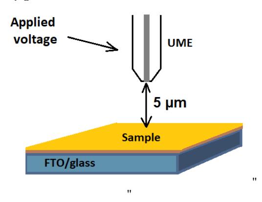
The scheme of the electroporation process is visualized in figure 1. Using SECM feedback mode, the ultramicroelectrode is approached to the sample. Then the electroporation process is being proceeded. Impedance spectroscopy is used to evaluate electrochemical changes of live yeast cells before and after electroporation. Using SECM scanning mode, surface changes are visualized after the electroporation. It was determined that a pore has been formed exactly below the working electrode.
Electroporation is already being used in various fields, such as medicine, gene engineering, or the food industry. Although the process is highly applicable, it is still random and incomplete controllable [5]. So, the goal of these studies is to develop a targeted electroporation process model, with good efficiency and easy performance.