In recent years, silver nanoparticles have been intensely studied for their unique optical, electrical, catalytic, thermal, and other related properties. Precious metal nanoparticles of a certain size, shape, and coatings amplify Raman signals from different analytical targets. This promising method is called shell-isolated nanoparticle-enhanced Raman spectroscopy (SHINERS) [1].
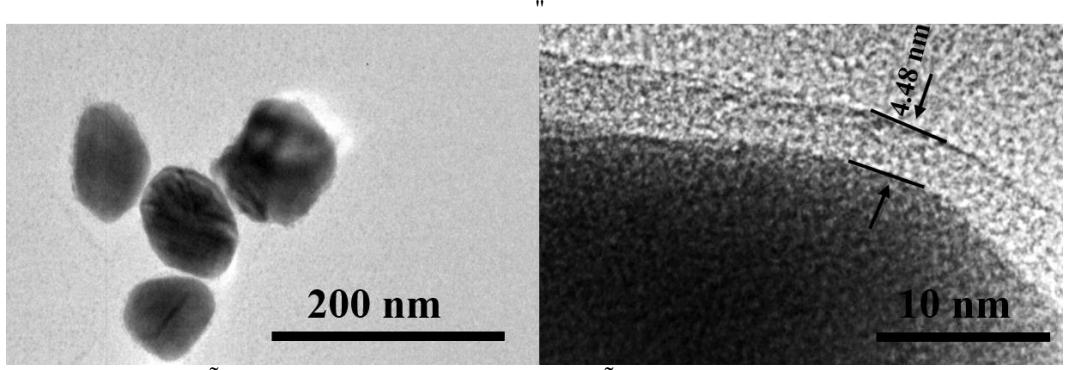
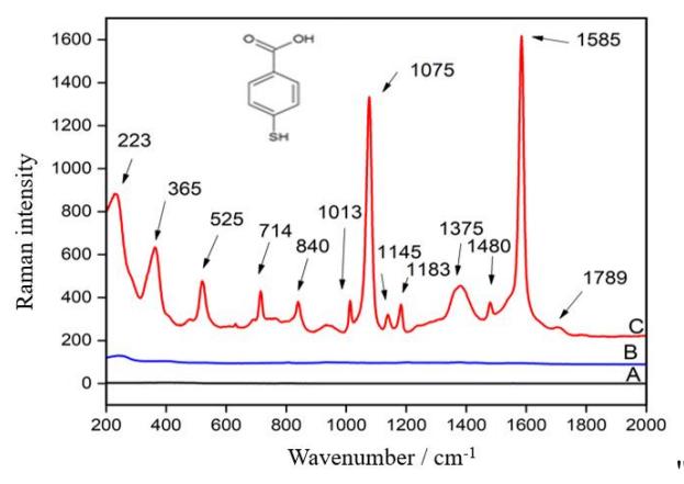
Various reducing and stabilizing agents are used to optimize the properties of synthesized nanoparticles [2]. Polyols are widely used because they help to maintain high temperatures and keep uniform heating. Microwave-assisted synthesis of nanostructures has several advantages over other methods: shorter reaction times, lower energy consumption, higher product yields with narrower size distributions [3]. Consequently, we combined microwave synthesis and chemical reduction to synthesize silver nanoparticles using popular polyols: ethylene glycol (EG), tetraethylene glycol (TEG), pentaethylene glycol (PEG), polyethylene glycol 200 (PEG-200). We aim to compare which agents are most suitable for obtaining stable, monodisperse silver nanoparticles. The nanoparticles meeting our criteria were coated with a silica (SiO2) shell to avoid direct chemical and electrical contact of the nanoparticles with a gold substrate, analyte, and environment. In this way, silver core-silica shell (Ag@SiO2) nanospheres with a size of 70±5 nm were synthesized (Fig. 1). A self-assembled monolayer of 4-mercaptobenzoic acid (MBA) was formed onto a gold plate and Ag@SiO2 nanoparticles were spread on it to amplify the Raman signal. A clear enhanced SHINERS spectrum of MBA on a smooth gold plate was obtained (Fig. 2).