
The manipulation of biological tissues, including their growth or regeneration is a very promising field. However, most of the current researches are in vivo or based on the biological tissue or organ sample donations that in most cases causes a discomfort for the patient. Therefore the development of micro/nano bioscaffolds is a successful alternative for the manipulation and growth of the biological samples in vitro.
In this study we analyse two types of commercial stereolithography 3D printers: Ember (Autodesk) and Asiga PICO 2 UV DLP and their abilities for printing bioscaffolds. The concept is based on the articles by Grigalevičiūtė [1] and Hart [2]. The authors of these articles present studies of the biocompatibility of printed structures with cells in the case of various widely used resins. It is also well known that bio-based resin can be used in 3D printing [3, 4] so there is a possibility to print any wanted biocompatible 3D geometry with a resolution of a few nanometers and this leads to accurate organs engineering. Methods for increasing biocompatibility in the following ways are also discussed: treatment with IPA (isopropyl alcohol), UV, thermal baking, autoclaving and coating with another material. All these steps are shown in Fig. 1
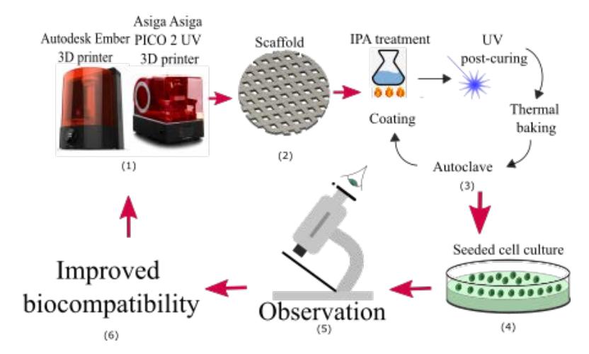
Based on the results obtained in previously mentioned articles it can be seen that Formlabs Clear and Flexible resins showed the best biocompatibility results. Taking into account the results obtained by Grigalevičiūtė, it is promising to achieve even better compatibility by changing the geometrical parameters of the scaffold (pore size, shape and porosity). Since cells die mostly in the middle of the scaffolds due to the pure leaching of monomers, changing of their geometry (making it with a higher resolution or creating a different geometric structures in the middle compared to the edges of the scaffold) might lead to even more biocompatible scaffolds. Various post-processing methods have also shown that the use of PDMS coating alone has greatly increased a biocompatibility. It would also be useful to try coating with other materials or even with several of them.