Development and spread of cancer are known to cause changes in the structure of extracellular matrix (ECM). A substantial portion of ECM is comprised of collagen, which is a noncentrosymmetric structure. Due to this, it is known to produce second-harmonic generation (SHG) signals. Furthermore, the SHG signals, produced in collagen, are strongly dependent on the angle between the collagen fibers and the incoming light polarization, hence the structural changes of collagen can be studied using polarimetric second-harmonic generation microscopy [1].
A reduced polarimetric SHG microscopy techniques, namely linear polarization-in, polarization-out (PIPO) microscopy, is applied for skin tissue imaging. In PIPO case, for each orientation of linearly polarized incoming laser radiation a set of linearly polarized states of outgoing second harmonic signal is measured [2].
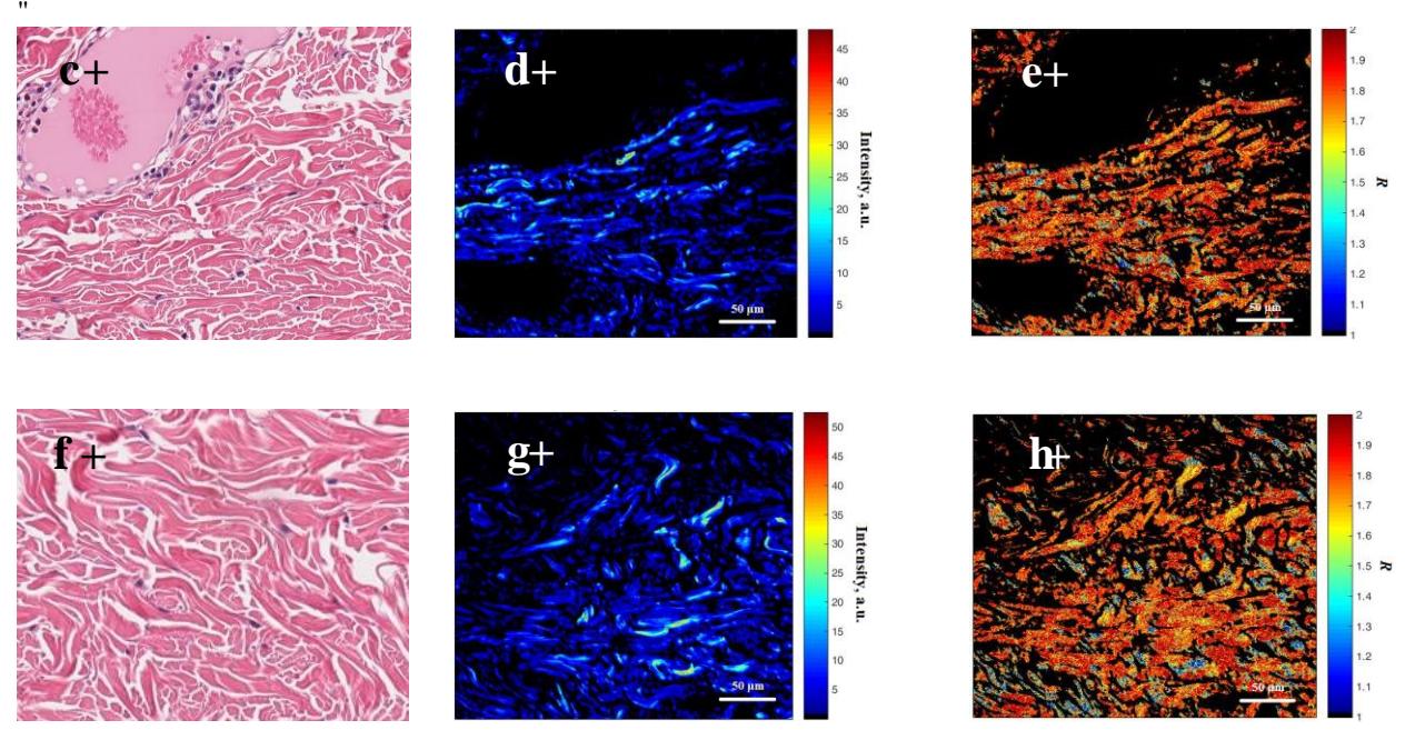
In this work, PIPO technique was applied to obtain the intensity, achiral susceptibility ratio $R$ and collagen fiber orientation angle distributions in pT1b stage lentiginous melanoma histological sections. The parameter distributions were extracted by performing a pixel by pixel fitting procedure of the intensity variations in the image using the SHG intensity equation for the PIPO case [2]. The same sample contained normal (intersection between healthy epidermis and dermis) and cancerous tissue (intersection between tumor tissue and dermis), therefore these parameters were used to investigate the structural differences between the two cases.
PIPO data analysis revealed the differences in collagen fiber orientations as well as increase in $R$ ratio and decrease in SHG intensity in cancerous tissues, which suggests a modified collagen structure. Therefore, the differences in the polarimetric parameters of collagen can be applied for melanoma research and diagnostics.