Cells produce a various heat shock proteins in response to stress. 70-kDa heat shock protein (Hsp70) is one of the best known heat shock proteins. Hsp70 participates in folding and assembly of newly synthesized proteins, refolding of misfolded and aggregated protiens. Hsp70 expresion is increased during stress conditions, and their function is vital for the recovery of cells after the insult [1]. Hsp70 is mainly localized in subcellular compartments, for example, cytosol, endoplasmic reticulum and mitochondria. In addition, Hsp70 is detected in extracellular space where this protein appear to play a different function from their chaperone activity [2].
Extracellular Hsp70 do not contain a consensus signal for their secretion via classical ER-Golgi compartment. Therefore, they are likely export by an alternative mechanism but the exact Hsp70 translocation mechanism across the plasma membrane remain to be elucidates.
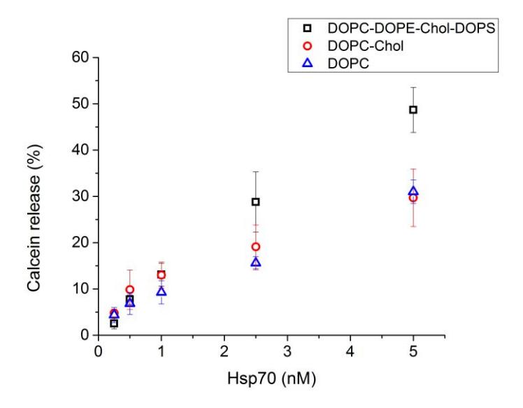
The aim of this work was to investigate Hsp70 interaction with various compositions unilamellar vesicles. In this study, Hsp70 was added to DOPC, DOPC/Chol (60:40) and DOPC/DOPE/Chol/DOPS (20:30:20:30) vesicles solutions and the change in fluorescence intensity due to calceine release from vesicles was detected. Results show that the incubation time of Hsp70 and unilamellar vesicles was 10 min. The smallest Hsp70 concentration which disturbs the integrity of membrane was 0,25 nM. Furthermore, fluorescence intensity of calcein release was about 1.5 times bigger at higher (>2nM) Hsp70 concentrations and vesicles contained negatively charged phospholipids (Fig. 1)