Conformational changes in proteins and their aggregation in the form of amyloid fibrils have been implicated in many neurodegenerative disorders, such as Alzheimer's, Parkinson's diseases or mammalian spongiform encephalopathy [1]. Currently, there is a lot of research carried out in order to examine how aggregation of amyloid proteins occurs and how various environmental factors may influence the processes of protein aggregation. It is observed that temperature, pH, ionic strength and type of shaking change not only the kinetic parameters of aggregation, but also the conformational structure of the protein, thus forming fibrils of different strains [2,3]. The fluorescent dye thioflavin-T is one of the most widely used dyes in amyloid protein studies [4]. In this work, it was investigated whether ionic strength of the solution affects the binding of thioflavin-T to amyloid fibrils using absorbance and fluorescence spectroscopy.
Insulin (200 μM protein dissolved in 100 mM NaCl 100 mM phosphate buffer (pH 2.4), incubation 24 h, 60 °C), lysozyme (200 μM protein dissolved in 2 M GuHCl 50 mM phosphate buffer (pH 6), incubation 24 h, 60 °C, with shaking, using glass beads), mouse prion-protein (0.5 mg/ml protein dissolved in 0.5 M GuHCl 50 mM phosphate buffer (pH 6), incubation 24 h, 60 °C, α-synuclein (200 μM protein dissolved in PBS, incubation 24h, 60 °C, with shaking, using glass beads) amyloid fibrils were prepared. After aggregation, the fibrils were centrifuged, resuspended into distilled water and concentrated to 400 μM). The absorbance of the samples was measured after mixing the fibrils, ThT and NaCl solutions (final protein conc. 100 μM) in the range from 300 to 600 nm. Excitation-emission matrices (EEM) of each sample were then scanned using an excitation and emission range from 435 to 500 nm. The samples were then centrifuged for 10 min at 9000 rpm and the supernatant absorbance in the range from 300 to 600 nm was measured.
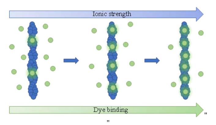
A direct dependence between the amount of ThT molecules bound to fibrils and the solution's ionic strength was observed. At higher ionic strength, more dye molecules are attached to the amyloid fibrils, which increases the sample fluorescence intensity (Fig. 1). Similar ionic strength effects were observed using all four amyloid proteins.