Laccases are multi-copper oxidases (EC 1.10.3.2), containing T1, T2 and T3 copper sites. The catalytic mechanism consists of several stages: i) the transfer of one electron and proton from the substrate to T1 Cu (oxidation); ii) the transfer of one electron from T1 to T2/T3 Cu cluster; iii) the T2/T3 cluster reduces one oxygen molecule to two water molecules, by using four electrons and protons. The reaction by product is water, thus this enzyme has high potential for industrial application [1]. Their natural substrates are aromatic compounds containing at least one hydroxyl, thiol, primary or secondary amine functional group.
Thought the years of investigating various laccases, a lot of information has already been accumulated: redox potentials, enzyme sources, reorganization energies, catalytic mechanism, etc. However, tangible application of laccases is hindered by the unresolved drawbacks such as poor stability, commercial unavailability, lack of efficient expression systems, low immobilization yields, etc.
One of the shortcomings for discovering laccases with new and/or novel features is a lack of substrates suitable for high-throughput screening and functional analysis. Currently, the most common compounds used for laccase functional analysis are 2,2'-azino-bis(3-ethylbenzothiazoline-6-sulphonic acid) under the trivial name ABTS and 4-[[2-[(3,5-dimethoxy-4-oxocyclohexa-2,5-dien-1-ylidene)methyl]hydrazinyl]methylidene]-2,6-dimethoxycyclohexa-2,5-dien-1-one known as syringaldazine [2,3]. Spectrophotometric activity assays with these substrates give acceptable results, but these compounds have poor stability, low specificity and are rather expensive.
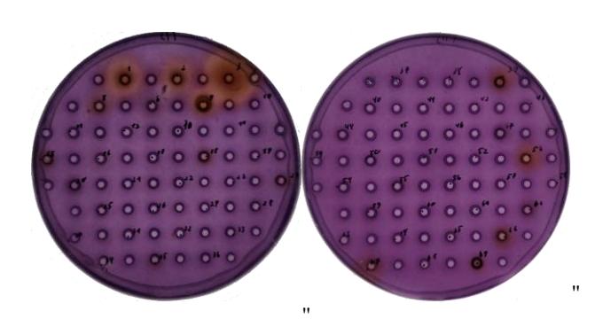
By our investigation, we present a new substrate for laccase activity screening on high-throughput agar-plates. This compound was synthesized using relevantly low priced aromatic diamine – N,N-dimethyl-p-phenylenediamine dihydrochloride. The latter substrate was tested on substrate-agar plates with commercially available laccase Novozym 51003 from Aspergillus oryzea (Fig. 1) and with the collection of wild fungi protein extracts provided by Jožef Stefan Institute (Fig. 2). The results in more detail will be presented during the poster session.