
Electrical impulses in plants can code and transfer information about various environmental changes and condition an appropriate response [1]. Generation of these impulses in plant cells heavily depends on the plasma membrane selective permeability to particular ions and their gradient on both sides of the membrane. Usually, Ca2+ concentration in the cytosol is kept low, whereas its increase causes the depolarization of the membrane potential and initiates action potential generation [2]. However, not much is known about the transmission characteristics of these impulses such as their propagation velocity and the compounds potentially affecting them.
We used an extracellular recording system [3] by placing an oblong cylinder-shaped Nitellopsis obtusa giant internodal cell along a chamber with electrically isolated compartments and fixing two potential-difference-recording electrode pairs. Knowing the exact distance between these pairs and measuring the time a potential took to propagate from one pair to another ($ \Delta t $, see Fig. 1), velocity of an action potential propagation can be evaluated. Experiments were performed using two types of stimuli in 10 min. intervals – electrical and of a low temperature solution, and two types of medium – artificial pond water (APW) and a solution of Ca2+ channel blocker verapamil (0.1 mM) a compound that might affect the impulse propagation velocity.
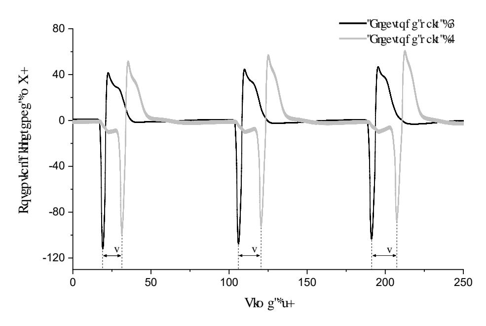
Results showed the dynamics of action potential propagation velocity in time: in control solution, regardless of stimulus modality, the very first stimulated potential propagated with the greatest velocity and with each given stimulus it decreased until the value steadied. 0.1 mM verapamil solution evoked a sequence of spontaneous potentials even before the stimulus could be provided. After the spontaneous activity ceased, we observed that the first stimulated action potential propagated the slowest with the velocity similar to the steadied one in the control group. Verapamil did not affect the action potential propagation velocity, but rather provoked spontaneous potentials and led the cell to death. The mortality of cells exposed to 0.1 mM verapamil proved to be of a 100%.
Hence, the action potential in plant cells propagates regardless of whether it is stimulated or spontaneous. The velocity of stimulated action potential propagation depends on if and how many times it has been recently evoked.